How do motion-compensated MRI techniques affect visualization of a moving object for RT planning?
Astrid van Lier,
The Netherlands
PD-0664
Abstract
How do motion-compensated MRI techniques affect visualization of a moving object for RT planning?
Authors: Astrid van Lier1, Pim Borman1, Martin Fast1, Bas Raaijmakers1
1UMC Utrecht, Radiotherapy, Utrecht, The Netherlands
Show Affiliations
Hide Affiliations
Purpose or Objective
Compressed SENSE (CS) and 3D VANE are MR techniques which recently became available for the 1.5 T Unity MR-linac (Elekta AB, Sweden). While 3D VANE scans are aimed at reducing motion artifacts by sharing k-space data between acquisitions, CS is used to decrease scan times by sparsely sampling k-space. In this phantom study we investigate the effect of using these techniques in relation to imaging moving structures under free breathing conditions for radiotherapy treatment planning.
Material and Methods
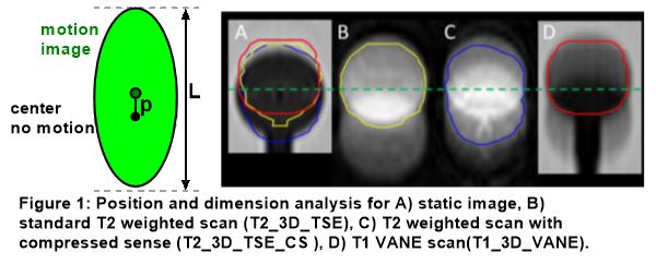
A 4D phantom (ModusQA, Canada) with a moving insert containing a 30 mm spherical target was used. Imaging was performed with and without motion. The motion pattern was cos4-shaped with a peak-to-peak amplitude of 20 mm and period of 4 seconds, oriented in cranial/caudal (CC) direction. Three types of scans were tested (T2_3D_TSE: SENSE factor 3; T2_3D_TSE_CS: CS factor 5; T1_3D_VANE XD Dixon: water image) with a reconstructed voxel of 0.64x0.64x2/0.64x0.64x2/0.78x0.78x1.5mm3 and a scan duration of 4:04/2:52/5:43 minutes, respectively. The spherical target was delineated. From the delineations the cranial/caudal (CC) length was extracted, which is a surrogate to measure if the GTV (30 mm), the ITV (30+20 = 50 mm) or a volume in between is shown. In addition, the center-of-volume (CoV) of the delineation was determined. A high and low contrast imprint of the sphere was visible in all motion images therefore a full (automatic using threshold) and tight (most prominent imprint of the spherical sphere only) delineation was performed (figure 1).
Results
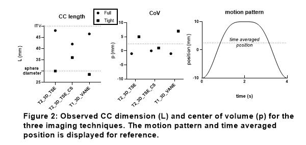
Static scans confirmed a CC sphere diameter of 30 mm for all 3 imaging techniques. For the motion scans, this full delineation diameter increased to 48/42/46.5 mm for T2 TSE/T2 TSE CS/VANE respectively (figure 2). The CoV deviated -1/0/-1 mm from the time-averaged (mid-)position. For the tight delineation a CC diameter of 30/36/28.5 mm was observed, while the CoV shifted to 5/1/7 mm compared to mid-position.
Conclusion
The observed CC diameter is largest in T2_3D_TSE and T1_3D_VANE and coincides with the definition of the ITV, while for T2_3D_TSE_CS a markedly decreased dimension was observed. CoV positions were close to mid-position for all techniques. For the tight delineation, the CC diameter of T2_3D_TSE and T1_3D_VANE were close to the actual sphere , however at the largest deviations from mid-position; especially T1_3D_VANE is positioned closer to end-expiration position. This end-expiration position is the most prevalent position (but not the time average position) in the cos4 signal which therefore prevailed in the T1_3D_VANE reconstruction. For CS reconstruction (T2_3D_TSE_CS), motion artifacts seem to be partly suppressed by the iterative reconstruction process which removes the non-coherent artifacts arising from sparse sampling.
Alterations in volume and position due to MRI acquisition/reconstruction techniques need to be considered in treatment planning and delivery.