Use of 4DMRI acquired on an MR-Linac to quantify intra-fraction motion in pancreatic cancer
Jessica Gough,
United Kingdom
PD-0658
Abstract
Use of 4DMRI acquired on an MR-Linac to quantify intra-fraction motion in pancreatic cancer
Authors: Jessica Gough1,2, Andreas Wetscherek2, Bastien Lecoeur2, Sophie Alexander1,2, Rosalyne Westley1,2, Brian Ng-Cheng-Hin1, Arabella Hunt1, Katharine Aitken1,2
1Royal Marsden Hospital, Radiotherapy, Sutton, United Kingdom; 2Institute of Cancer Research, Radiotherapy and Imaging, London, United Kingdom
Show Affiliations
Hide Affiliations
Purpose or Objective
Dose-escalated radiotherapy (RT) may improve survival for patients with locally advanced pancreatic cancer (LAPC)(1,2). However, accuracy of delivery can be compromised by intra-fraction motion of the target and adjacent organs at risk (OARs). MR Linacs (MRLs) are a novel platform for LAPC stereotactic body RT (SBRT) delivery, maximising accuracy through use of improved image guidance and adaptive planning thus enabling dose escalation without exceeding OAR tolerance. Abdominal compression (AC) may be used for SBRT where more advanced motion management methods e.g. tracking are not available. Current workflows utilise 4DCT to quantify residual motion and determine internal target volumes (ITVs). However, 4DCTs are acquired once prior to treatment and no further 4D imaging is routinely acquired, assuming that motion remains unchanged. This study evaluated use of 4DMRI, acquired on an Elekta Unity MR-Linac (Elekta AB, Stockholm, Sweden), to quantify intra-fraction target and OAR motion and the effects of AC.
Material and Methods
Patients with LAPC undergoing RT on standard linear accelerators were recruited to undergo MRL imaging under the PRIMER (NCT02973828) study. Patients underwent scans with and without AC with a ZifixTM (Qfix,USA) belt, including balanced 3DVane sequences acquired in free breathing. The 3DVane is a volumetric radial gradient echo sequence with stack-of-stars readout and golden-angle spacing. 3DVanes were retrospectively reconstructed into 4DMRI comprised of 10 respiratory phases using an in-house implementation of XD-GRASP(3). Pancreatic gross target volumes (GTVs) and OARs were delineated on maximum inspiration and expiration 4DMRI phases. Diaphragmatic motion on 4DMRI was compared with corresponding 4DCTs without AC for each patient. Centre of mass (COM) motion coordinates of GTVs and OARs were extracted to calculate peak to peak motion in all planes and compared with and without AC.
Results
10 LAPC patients were recruited and 44 3DVane sequences acquired during 22 scanning sessions. Some were not possible to reconstruct due to data corruption or missing raw data (n=8). 36 4DMRI were analysed: without AC (n=18), with AC (n=18). There was no significant difference in diaphragmatic motion between 4DCT and 4DMRI (median 11.4mm (range 4.1-28.9mm) vs. median 10.4mm (range 4.3-22.7mm) respectively, p =0.082). Target and OAR motion on 4DMRI was greatest in the superior-inferior plane and there was a reduction in motion with AC. Median superior-inferior COM motion with and without AC of GTV and OARs are shown in Table 1.
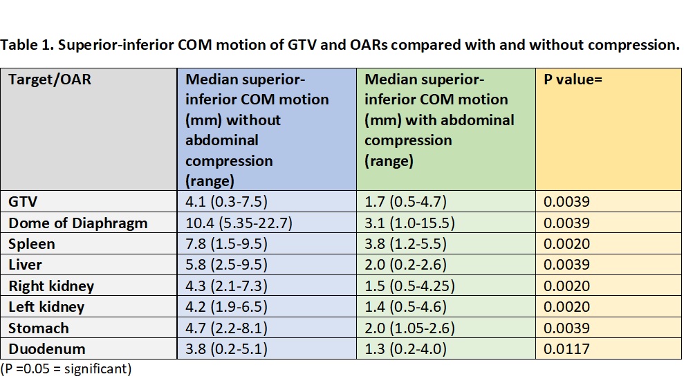
Conclusion
It is feasible to quantify intra-fraction motion using 4DMRI (reconstructed from 3DVane) acquired on an MRL and this is reduced with AC. 4DMRI acquired on treatment days could be used to quantify daily intra-fraction GTV motion and verify margin expansions. These data could potentially be used to better understand intra-fraction dose deposition and inform development of advanced motion compensation strategies for abdominal RT on the MRL.