Session Item

Monday
May 09
08:00 - 08:40
Room D1
Modern imaging in radiation oncology
Esther Troost, Germany
3010
Teaching lecture
Clinical
11:25 - 11:35
Dosimetric evaluation of OAR in APBI patients treated with multicatheter interstitial brachytherapy
OC-0015

Abstract

Dosimetric evaluation of OAR in APBI patients treated with multicatheter interstitial brachytherapy
Authors:

Marina Macaes1, Sara Pinto1, Alexandre Pereira1, Joana Lencart1, Pedro Fernandes2, Lurdes Trigo2

1Portuguese Oncology Institute of Porto , Medical Physics, Porto, Portugal; 2Portuguese Oncology Institute of Porto , Brachytherapy Service, Porto, Portugal

Show Affiliations
Purpose or Objective

Dosimetric evaluation of organs at risk (OARs) according to the recent GEC-ESTRO recommendations (1) in sixty-one APBI patients treated with multicatheter interstitial brachytherapy (MIBT) using a HDR Ir-192 source.

Material and Methods

Between February 2017 and December 2019, 61 APBI patients (32 patients (52%) with left-sided and 29 (48%) with right-sided breast tumors) were treated with MIBT technique, with a prescription dose of 32Gy (4Gy/fraction in 5 days with a minimum interval between fractions of six hours). After lumpectomy, a post-operative multicatheter implant was performed. After catheter insertion, the patients underwent a CT scan (2mm slices), and treatment plans were obtained with TPS Oncentra MasterPlan v4.1. The target volumes and the OARs, including skin, lung ipsilateral, ipsilateral non-target breast and heart (in the cases of left-sided tumors) were delineated and the dose-volume histograms (DVH) were evaluated.

Results

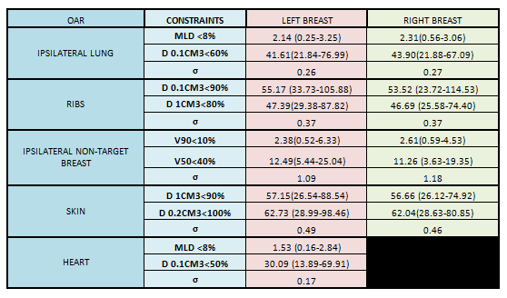
Regarding the tumor location in breast, we did not find a relation between the quadrant and the dose received by OAR. Table 1 shows the analyzed dose-volume parameters and the recommended limits.


The mean values in all the parameters were below the limits of GEC-ESTRO recommendations, and doses to skin and ipsilateral non-target breast were low for all patients. However, some patient’s heart, ribs and ipsilateral lung dose exceeded the constraints.


The minimum and maximum heart-to-PTV distance was 2mm and 46mm, respectively, and cardiac dose increased with decreased heart-to-PTV distance.


Regarding the ribs the average D0.1cm3 and D1cm3 values were 53.89% and 46.84%, respectively, but 5 patients exceeded dose-volume parameters because of PTV-ribs proximity.

Of the 61 patients, 9 exceeded the dose-volume parameter D0.1cm3 <60% to lung. Evaluating those cases, we detected that all of them had the 50% isodose curve reaching the lung, opposing the rest of the patients. 


Conclusion

The average of all dose-volume parameters was in conformity with guidelines, however there were patients (16.3%) that exceeded some recommendations because of PTV’s location relative to OAR, mainly lung, ribs and heart. It is important to take it in consideration the follow-up of patients regarding toxicity.

APBI using multicatheter brachytherapy can reduce radiation exposure of organs-at-risk, nevertheless a anatomic tumor position study, such as distance from heart to lumpectomy bed, can help in individualized technique selection that ensure the lowest dose possible at OAR.