Vienna, Austria

ESTRO 2023

Session Item

Microenvironment
8005
Poster (Digital)
Radiobiology
The inflammatory environment analysis of acute radiation enteritis for cervical cancer
Chenying Ma, China
PO-2208

Abstract

The inflammatory environment analysis of acute radiation enteritis for cervical cancer
Authors:

Chenying Ma1, Jing Zhao1, Xiao-ting Xu1, Xiao-ping Li2, Xiao-lan He3, Mu-xing Zhang4, Yan-zheng Gu5, Ju-ying Zhou6

1the First Affiliated Hospital of Soochow University Suzhou, Department of Radiation Oncology, Suzhou, China; 2the First Affiliated Hospital of Soochow University, Department of Critical Care Medicine, Suzhou, China; 3the First Affiliated Hospital of Soochow University, Department of Radiation Oncology, Suzhou, China; 4the First Affiliated Hospital of Soochow University, Department of Clinical Nutrition, Suzhou, China; 5Soochow University, Jiangsu Key Laboratory of Clinical Immunology, , Suzhou, China; 6 the First Affiliated Hospital of Soochow University, Department of Radiation Oncology, Suzhou, China

Show Affiliations
Purpose or Objective

To identify the immune characteristic phenotypes of the gut injury in radiation enteritis (RE) and severe acute radiation enteritis (SARE) and to determine the classification pattern of gut microbiota symbionts on immunity from the perspective of omics analysis and molecular signaling pathways through the inflammatory environmental factors in peripheral blood.

Material and Methods

The secretion of serum IL-2, IL-4, IL-6, IL-10, TNF, IFN-γ and IL-17a proteins in cervical cancer VMAT patients during radiotherapy were detected by Cytometric Bead Array (CBA) technology. The secretion of FOXP3, COX-2, TLRs, PD-1, PD-L1, EGFR and MAPK proteins were detected by Enzyme-Linked Immunosorbent Assays (ELISA). A co-culture system of fecal suspension from the cervical cancer patients during VMAT and human colorectal mucosal cells (FHC) cells were constructed. The protein expressions of Iκκ, p-Iκκ, NF-κB-p65, IL-6 and COX-2 were detected by Western blot (WB).

Results

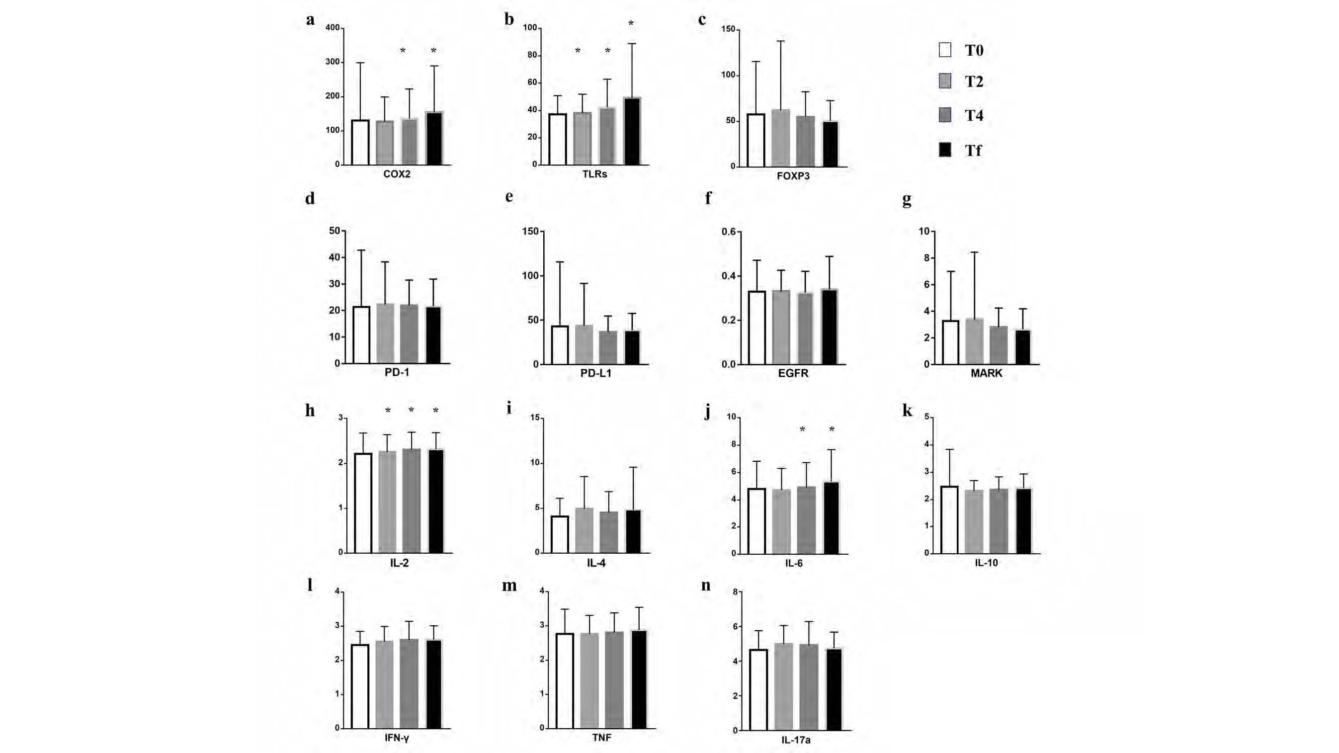
1. It was found that in addition to IL-10, COX-2, TLRs, FOXP3, PD-1, PD-L1, EGFR, MAPK, IL-2, IL-4, IL-6, IFN-γ, TNF, and IL-17a molecules increased during VMAT compared with baseline through the determination of inflammatory factor proteins in peripheral blood, among which the secretion of COX-2, TLRs, IL-2 and IL-6 were time/dose dependent.


2. Through ROC curve analysis, it was found that IL-6 could be used as an inflammatory molecule to predict RE, and IL-6 and COX-2 could be used as inflammatory molecules to predict SARE. The inflammatory factors of RE and SARE were preliminarily identified.
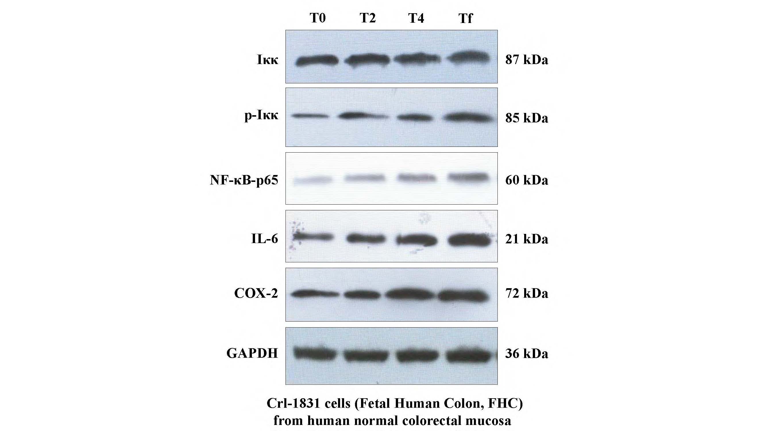
3. The supernatant and cell lysate which were co-cultured with the fecal suspension from cervical cancer patients during VMAT and FHC were collected for total protein extraction. The protein expressions of p-Iκκ, NF-κB-p65, IL-6 and COX-2 were found to be time/dose dependent. Moreover, the phosphorylation of Iκκβ at Ser176 and Ser177 up-regulated the expression of NF-κB-p65, IL-6 and COX-2 downstream of the pathway.


Conclusion

1. The TLR signaling pathway was activated via gut microbiota after radiation. And the secretion of downstream IL-6 and COX-2 increased with radiation dose, both of which could be the inflammatory environmental factors to predict RE and SARE.
2. It was verified by WB experiments that the symbiotic environment of gut microbiota affected by radiation was activating by the classical NF-κB pathway, which followed by up-regulation of downstream pro-inflammatory molecules.