Vienna, Austria

ESTRO 2023

Session Item

Sunday
May 14
09:00 - 10:00
Business Suite 3-4
Clinical Brachytherapy
Alina Sturdza, Austria
2190
Poster Discussion
Brachytherapy
Effects of simulated systematic applicator displacement in intra-cavitary HDR cervical brachytherapy
Orla Houlihan, United Kingdom
PD-0408

Abstract

Effects of simulated systematic applicator displacement in intra-cavitary HDR cervical brachytherapy
Authors:

Orla Houlihan1,2, Monica Byrne3, Owen McLaughlin3,4, Geraldine Workman3, Sergio Esteve3, Ursula McGivern1, Anne Drake1, Elizabeth Baird1, Kevin Prise2, Alan Hounsell3, Suneil Jain1,2

1Northern Ireland Cancer Centre, Belfast Health and Social Care Trust, Clinical Oncology, Belfast, United Kingdom; 2Queen's University Belfast, Patrick G. Johnston Centre for Cancer Research, Belfast, United Kingdom; 3Northern Ireland Cancer Centre, Belfast Health and Social Care Trust, Radiotherapy Medical Physics, Belfast, United Kingdom; 4Patrick G. Johnston Centre for Cancer Research, Queen's University Belfast, Belfast, United Kingdom

Show Affiliations
Purpose or Objective

To simulate systematic applicator displacements in intra-cavitary HDR cervical brachytherapy with the view to determining the required accuracy of in vivo dosimeters developed as part of the EU H2020 ORIGIN project.

Material and Methods

20 CTs performed prior to the first fraction of intra-cavitary HDR cervical brachytherapy were included. Organs at risk of interest were bladder, rectum and bowel. The bladder and rectum were divided into subsections of superior, middle and inferior along divisions of the upper, middle and lower third of the high risk clinical target volume (HRCTV)(Fig. 1). The ring and tandem applicator was systematically displaced in 1 mm increments from 1-6 mm in cranial, caudal, anterior, posterior, left and right directions along the plane of the central tandem to simulate applicator displacement between planning CT and brachytherapy delivery. The D2cc (minimum dose to the most irradiation 2cc) was calculated for each displacement and compared to the original planning CT.


Results

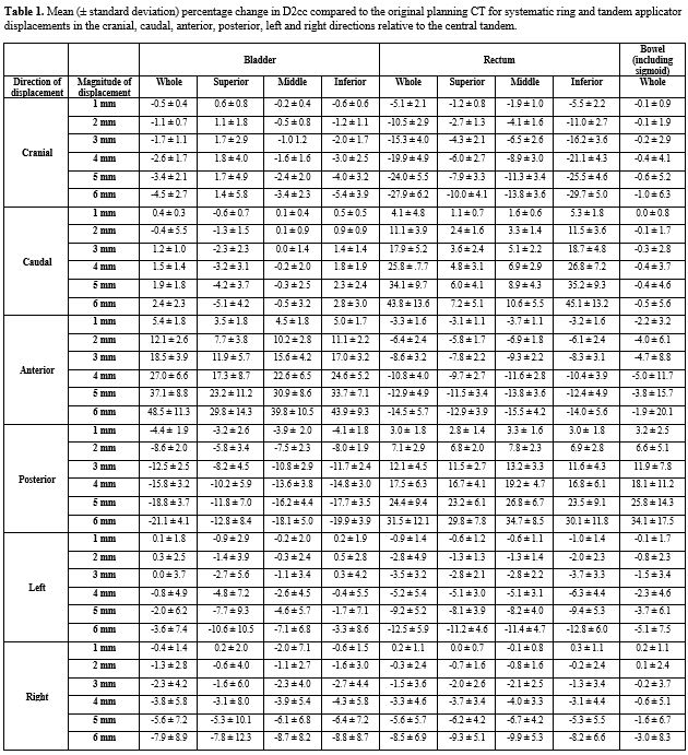
The greatest mean increases in D2cc occurred in the anterior direction for bladder and posterior direction for rectum, with increases of 5.4±1.8%, 18.5±3.9% and 37.1±8.8% to bladder and 3.0±1.8%, 12.1±4.5% and 24.4±9.4% to rectum for 1, 3 and 5 mm shifts in each respective direction. The greatest mean increases in bowel D2cc occurred with posterior displacement with increases of 3.2±2.5%, 11.9±7.8% and 25.8±14.3% for 1, 3 and 5 mm shifts respectively. 

The inferior bladder experienced the greatest percentage change in D2cc with 1, 3 and 5 mm anterior shifts resulting in mean increases of 5.0±1.7%, 17.0±3.2% and 33.7±7.1% respectively. The middle part of the rectum experienced the greatest percentage change in D2cc with 1, 3 and 5 mm posterior shifts resulting in mean increases of 3.3±1.6%, 13.2±3.3% and 26.8±6.7% respectively. 

Wide deviations in mean D2cc occurred with increasing displacement, with shifts of 6 mm resulting in changes of 48.5±11.3%, -21.1±4.1% and -7.9±8.9% in the anterior, posterior and right directions for the bladder, 27.9±6.2%, 43.8±13.6%, -14.5±5.7%, 31.5±12.1%, -12.5±5.9% and -8.5±6.9% in cranial, caudal, anterior, posterior, left and right directions for the rectum and 34.1±17.5% in the posterior direction for the bowel. Displacements in the caudal and left directions had the least mean changes for bladder at 2.4±2.3% and 3.6±7.4% at 6 mm respectively. Displacements in the cranial and caudal directions had the least mean changes for bowel at -1.0±6.3% and -0.5±5.6% at 6 mm respectively (Table 1).


Conclusion

The greatest mean increase in dose to bladder occurred with displacements anteriorly and to rectum and bowel posteriorly, in keeping with anatomical locations. Applicator displacements as small as 1 mm can result in increases in D2cc of >5% and dosimeters should have the ability to detect displacements at least as small as this. The effect of applicator displacements on bladder and rectum subsections is important for determining the optimal positions for dosimeters.