Method for regular calibration of small point detector in brachytherapy
Marjolein Heidotting,
Denmark
PD-0504
Abstract
Method for regular calibration of small point detector in brachytherapy
Authors: Marjolein L.E. Heidotting1, Peter Georgi1, Søren Kynde Nielsen2, Anders Traberg Hansen2, Harald Spejlborg2, Steffen Bjerre Hokland2, Kari Tanderup2, Jacob G. Johansen3
1Aarhus University, Department of Clinical Medicine, Aarhus, Denmark; 2Aarhus University, Department of Oncology, Aarhus, Denmark; 3Aarhus University, Danish Center for Particle Therapy, Aarhus, Denmark
Show Affiliations
Hide Affiliations
Purpose or Objective
Development of a simple method for routine recalibration of small point detectors used for in vivo dosimetry (IVD) during brachytherapy (BT).
Material and Methods
IVD is a standard part of BT treatments at our clinic. The IVD is performed using a small detector based on a scintillating crystal (1x0.5x0.5 mm^3) coupled to a Si-Diode through a 15 m long optical fiber. During BT, the probe is placed inside the target area through BT catheters and the dose rate is read out at 20 Hz. To protect the probe, the optical fiber is detached from the Si-diode after each treatment, and reattached just prior to the next treatment. The response of the detector varies as it is detached and attached. This gives rise to the need of recalibrating the detector before each use. A simple method, which takes <10 min and can be performed as part of the pre-treatment QA, has been developed.
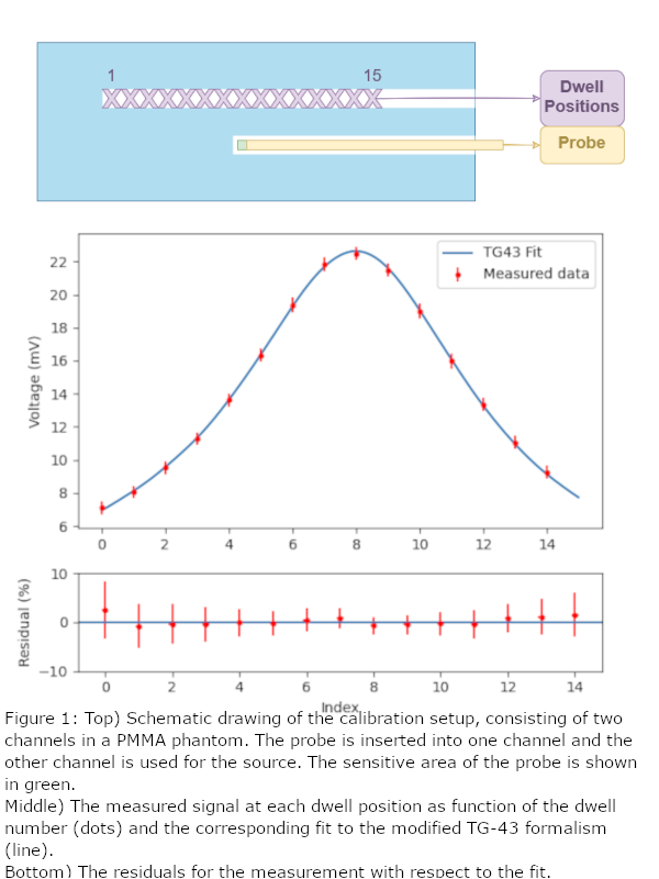
The calibration is performed using a 30x12x12 cm^3 phantom made from PMMA (fig. 1 (top)). In this phantom there are two straight and parallel needles placed 20 mm apart; one for the probe, which is placed at the bottom of the needle, and one which is attached to an afterloader. A measurement is performed with 15 dwell positions, spaced 5 mm apart, each with a fixed dwell time of 10 s. A function based on a modified version of the TG43 formalism is fitted to the 15 data points (fig. 1). The modifications account for the missing scatter in the phantom. The calibration factor (from voltage to dose rate) (CF) and the location relative to the bottom of the needle of the closest approach to the sensitive area of the probe (ΔZ) are used as fitting parameters. This procedure was performed on data from 48 different calibrations, using 4 different probes between March 2019 and December 2020.
Results
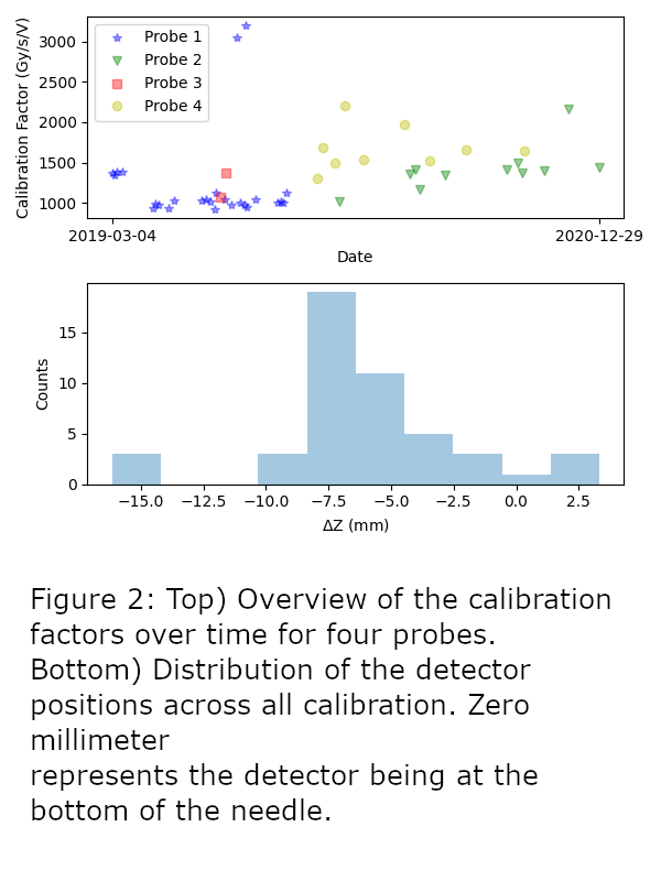
An example of a fit together with the residuals is shown in the middle of fig. 1. The mean reduced χ2 of all fits is 0.110 ± 0.097 (1SD). ΔZ varied between -15.2 and 3.3 mm from the expected location (fig. 2 (bottom)). Large fluctuations (up to 46% (1SD)) are seen in the calibration factors for each probe (fig. 2 (top)). This is expected to stem from the re-attachment of the probe. Furthermore, the calibration factor for probe 1 and 2 increased over time, which indicates degradation of the probe sensitivity.
Conclusion
We have presented a method to calibrate small point detectors, which is easily implementable and can be performed in less than 10 minutes. The method compensates for uncertainties in ΔZ, making it robust against positional uncertainties, which can lead to large calibration uncertainties, due to the steep BT dose gradients. The calibration factors vary both on the long and short term, based on both degradation of the material and coupling of the probe. This variation can be as large as a factor of three and confirms the need for regular recalibration.