A pre-clinical, phantom based study for real time source position monitoring in HDR brachytherapy
PD-0503
Abstract
A pre-clinical, phantom based study for real time source position monitoring in HDR brachytherapy
Authors: Aleyna Sipahi1, Johannes Strotmann2,3, Ndimofor Chofor4, Ioannis Simiantonakis1, Amol Patil5, Andreas A. Schönfeld4
1Heinrich Heine University, Department of Radiation Oncology, Duesseldorf, Germany; 2Carl von Ossietzky University, University Clinic of Medical Radiation Physics, Oldenburg, Germany; 3University Clinic of Medical Radiation Physics, Medical Campus Pius-Hospital, Oldenburg, Germany; 4Sun Nuclear, A Mirion Medical Company, Research & Development, Melbourne, USA; 5Mirion Technologies, Inc., Research & Development, Meriden, USA
Show Affiliations
Hide Affiliations
Purpose or Objective
In high dose rate brachytherapy, technical malfunctions of the equipment or unexpected complications during the therapeutic intervention can lead to misapplication of the dose and thus to severe consequences for the patient and personnel. In this project we performed a pre-clinical phantom investigation of a prototype gamma-camera system for real-time source localization under clinical circumstances.
Material and Methods
The prototype HDR-Vue© (Mirion Technologies, Atlanta, USA) consists of a gamma camera and an optical video camera. By co-registering both datasets in real-time, a source position indicator is superimposed onto the optical camera’s image. The system was tested using a CBCT electron density phantom (Sun Nuclear, A Mirion Medical Company, Norfolk, USA) to simulate attenuation and scatter within a patient. A stainless steel applicator probe of a GammaMedplus iX afterloader (Varian Medical Systems, Palo Alto, USA) was positioned inside the phantom and the phantom was placed in the center of the HDR-Vue's FOV at a source-to-camera distance of 50 cm. The Iridium-192 source had 20 stopping positions with dwell times of 5 seconds each and a step width of 5 mm.
Results
The HDR-Vue was able to visualize the real-time position of the Iridium-192 source during the radiation. A red circle in the live image indicates the localized real-time position of the radioactive source at any moment of the radiation. Figure 1 shows a composition of three images recorded when the source was at positions 1, 10 and 20.
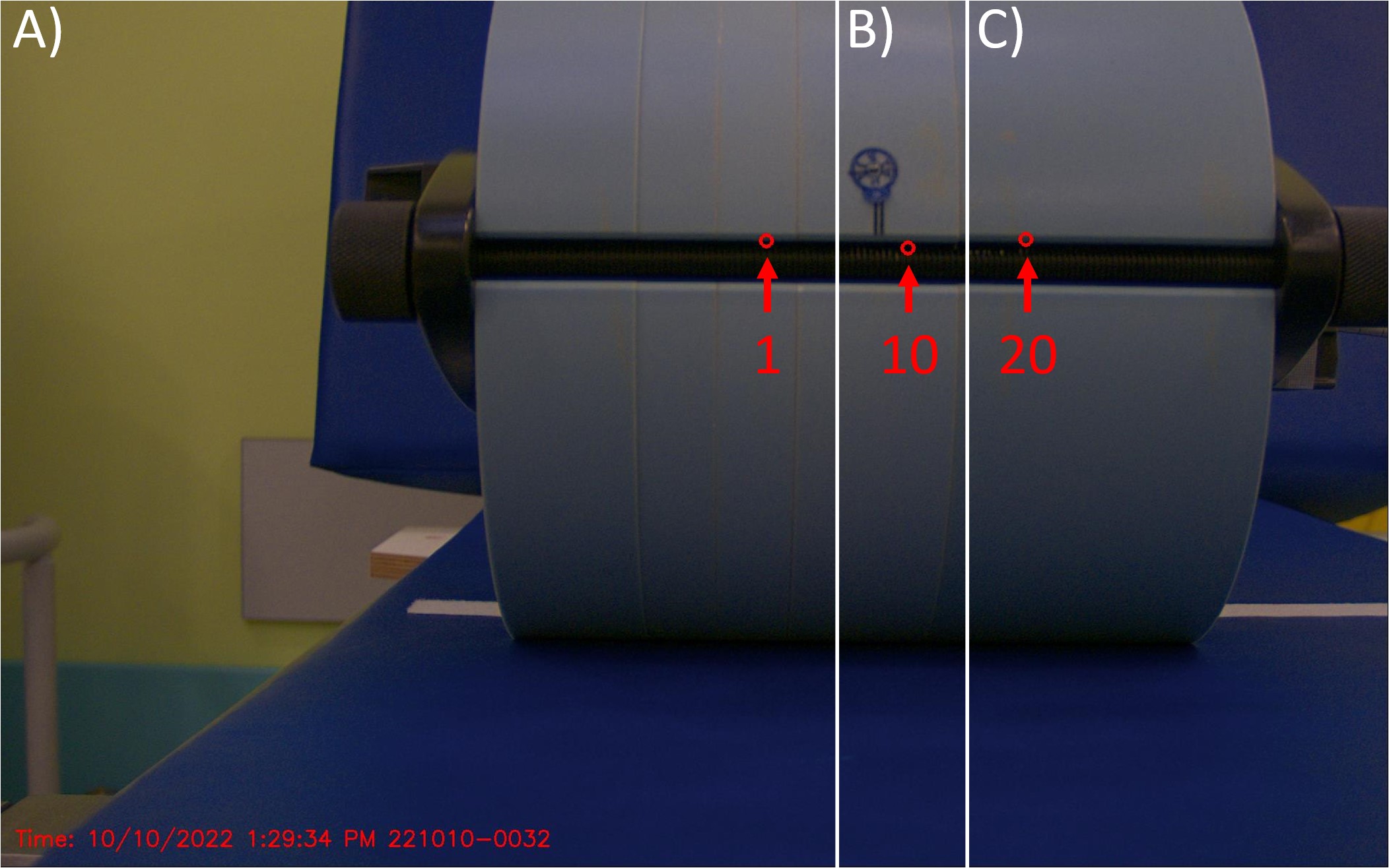
Figure 1: Overlay of three images showing different source locations as visualized by the HDR-Vue system. The Iridium-192 source was at dwell position 1 in section A) of the image, and in positions 10 and 20 in sections B) and C) respectively.
Conclusion
The measurements have shown that the HDR-Vue can detect the position of an Iridium-192 source in real-time, even when the source is inside a phantom. The ability to monitor the HDR source may help to detect gross errors and enhance treatment safety. Also, the knowledge of the source position facilitates patient evacuation in the event of incidents or accidents.