Vienna, Austria

ESTRO 2023

Session Item

Saturday
May 13
16:45 - 17:45
Business Suite 1-2
Technical improvements in radiotherapy practice
Rianne de Jong, The Netherlands
1630
Poster Discussion
RTT
Deep learning tumor segmentation for target delineation in glioblastoma using multi-parametric MRI
Marianne Hannisdal, Norway
PD-0316

Abstract

Deep learning tumor segmentation for target delineation in glioblastoma using multi-parametric MRI
Authors:

Marianne Hannisdal1, Dorota Goplen2, Saruar Alam3, Judit Haasz4, Leif Oltedal5, Mohummad Aminur Rahman6, Cecilie Brekke Rygh5, Stein Atle Lie7, Arvid Lundervold3, Martha Chekenya6

1Haukeland University Hospital, Dept of Oncology and Medical Physics, Bergen, Norway; 2Haukeland University Hospital, Dept of Oncology, Bergen, Norway; 3Mohn Medical Imaging and Visualization Centre, Radiology, Bergen, Norway; 4Haukland University Hospital, Radiology, Bergen, Norway; 5Haukeland University Hospital, Radiology, Bergen, Norway; 6University of Bergen, Biomedicine, Bergen, Norway; 7University of Bergen, Clinical Odontology, Bergen, Norway

Show Affiliations
Purpose or Objective

High precision tumor delineation is a prerequisite for optimal RT treatment planning that enables precise organ-at-risk sparing and reduction of adverse effects. However, manual tumor delineation remains laborious and challenging due to radiological complexity. The objective of this study was to investigate the feasibility of the HD-GLIO tool, an ensemble of pre-trained deep learning models based on the nnUNet algorithm, for glioblastoma tumor segmentation.

Material and Methods

In a collection of 29 multi-parametric standard MRI examinations from 13 glioblastoma patients, we compared the predicted contrast-enhanced (CE) and non-enhancing (NE) output volumes from HD-GLIO segmentation and the corresponding manual delineations obtained by two independent expert operators (Figure 1). 

The output of HD-GLIO was compared by Dice Similarity Coefficient and Hausdorff 95% (HD95) to: (i) the manual delineations of each observer separately, emphasizing differences across disciplines, (ii) the fused label, being the union and joint contribution from two disciplines to the manual delineations, and (iii) the aforementioned labels with added isotropic dilations, representing margins clinically used in RT. We also assessed the volume consistency between measures by inter-item intraclass correlation coefficient (ICC).


Results

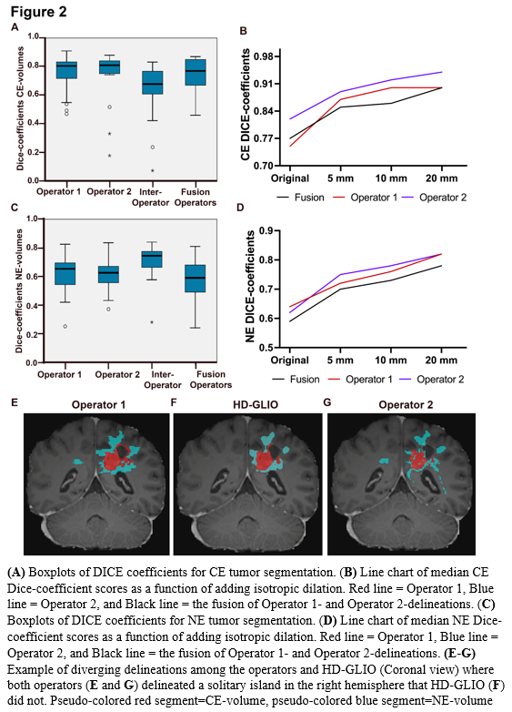
For CE, median Dice scores were 0.81 (95% CI 0.71-0.83) and 0.82 (95% CI 0.74-0.84), while median HD95 were 5.91 (95% CI 2.8-16.4) and 3.16 (95% CI 2.8-7.1) for Operator-1 and Operator-2, respectively (Figure 2 A). For NE, median Dice scores were 0.65 (95% CI 0.56-0,69) and 0.63 (95% CI 0.57-0.67), while median HD95 were 16.1 (95% CI 10.6-22.2) and 16.7 (95% CI 9.4-23.2), respectively (Figure 2 C).

Comparing volume sizes, we found excellent ICC of 0.90 (p<0.001) and 0.95 (p<0.001), for CE, respectively, and 0.97 (p<0.001) and 0.90 (p<0.001), for NE, respectively. Moreover, there was a strong Spearman’s correlation of 0.83 (p<0.001) between RANO-volumes and HD-GLIO-volumes.

Taken together, we found that for CE-volumes, the Dice similarity coefficients and HD95 had better scores between operator and HD-GLIO segmentation, than for inter-operator scores. This indicates that the HD-GLIO segments had a shape and location somewhat intermediate between the Operator-1 and Operator-2 manual delineations. Adding dilations further increased the Dice-scores and reduced the relative performance difference between individuals. For NE-volumes, we found that Dice similarity and HD95 showed poorer agreement between operator and HD-GLIO than inter-operator scores. This was largely because manual NE-delineations held substantially larger volumes than HD-GLIO predictions

Average processing time was < 6 minutes per dataset.


Conclusion

HD-GLIO deep learning predictions demonstrated high geometrical agreement with manual delineations in segmenting glioblastoma tumor compartments on standard multi-parametric MRI. We therefore find HD-GLIO feasible as an oncologist support tool for target delineation.