Development of an anthropomorphic 4D Phantom for multimodal imaging, 4D radiation and SGRT
Anahita Bakhtiari Moghaddam,
Germany
PO-2314
Abstract
Development of an anthropomorphic 4D Phantom for multimodal imaging, 4D radiation and SGRT
Authors: Anahita Bakhtiari Moghaddam1
1german cancer research center (dkfz), Medical Physics in Radiation Oncology, Heidelberg, Germany
Show Affiliations
Hide Affiliations
Purpose or Objective
inevitable patient movements during imaging can result in interfractional and intrafractional errors, originating mainly from the respiratory motions during the radiation planning, namely dosimetry planning. These errors might also arise from issues in patient positioning or be due to imaging artifacts.
Advancements in four-dimensional (4D) radiotherapy and the development of adaptive radiotherapy that examines patient motion during imaging and radiotherapy can facilitate the recognition and reduction of such errors. Use of Surface Guided Radiation Therapy (SGRT) makes it possible to get even closer to this goal. Using an anthropomorphic 4D phantom, the quality of radiation can be verified from start to the end.
Material and Methods
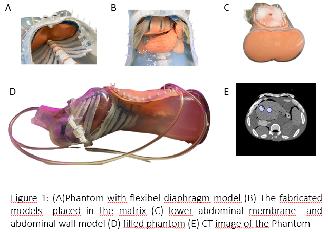
The phantom includes the following organ model: lungs, liver, kidneys, spleen, duodenum, stomach, pancreas, parts of the large and small intestine and bones of the spine.The required organs and bones were segmented using CT scan data of a patient and three-dimensional models were created. using CAD software, the mold for the organ and bone models can be designed. The models are produced using the 3D printer.
After fabricating the molds, depending on the requirements of the model, they were filled with either a prepared liquid contrast gel NiDTPA-KCl agarose mixture, a liquid contrast, or silicone.
Three tumor models were applied in the liver model, each of which was loaded with EBT3 measurement films.
The phantom body was made in a form of a mannequin from transparent acrylonitrile butadiene styrene (ABS). The fabricated models were placed in a matrix of superabsorbent and agarose gel.
Using a hydraulic system, the lung models were designed to be filled with air and the lower abdominal membrane can be pressed mechanically. The pressure generated by the diaphragm model in the abdominal region and the lower abdominal membrane pressed by an actuator make it possible to simulate a respiratory movement in the abdominal cavity. It also causes a movement in the abdominal wall model.
Results
The phantom simulates anatomically realistic organs and tumoral lesions and is also able to simulate human respiratory motion, which can be used for multimodal imaging. The models have anatomically equivalent radiographic scattering and absorption properties and produce realistic relation times in MR. The target surface of the phantom could be successfully acquired with the 3D cameras of the AlignRT.
Conclusion
This generated phantom can be used for radiation planning of the liver tumor models. In addition, real-time automated segmentation provided by this phantom makes it more feasible to perform an adaptive radiation planning and to do a quality assessment of respiratory-guided radiation, using AlignRT. However, reproducability of the organ movements is yet to be determined.
A place for the use of EBT3 Film imaging has been established in Phantom. Moreover, other detectors such as ionization chambers or dosimetric gels may be used for dose measurement in Phantom in the future.