Vienna, Austria

ESTRO 2023

Session Item

Optimisation, algorithms and applications for ion beam treatment planning
7008
Poster (Digital)
Physics
Monitoring and Quality Assurance of scanned proton pencil beams using a CMOS detector
Allison Toltz, United Kingdom
PO-1990

Abstract

Monitoring and Quality Assurance of scanned proton pencil beams using a CMOS detector
Authors:

Samuel Flynn1,2, Colin Baker3,4, Michael Homer1, Vasilis Rompokos3,4, Russell Thomas1,5, Allison Toltz3,4, Tony Price2,1

1National Physical Laboratory, Medical Radiation Science, Teddington, United Kingdom; 2University of Birmingham, Particle Physics, Birmingham, United Kingdom; 3University College London Hospitals NHS Foundation Trust, Department of Radiotherapy Physics, London, United Kingdom; 4University College London, Department of Medical Physics and Biomedical Engineering, London, United Kingdom; 5University of Surrey, Faculty of Engineering and Physical Sciences, Guildford, United Kingdom

Show Affiliations
Purpose or Objective

The effectiveness of Pencil Beam Scanning (PBS) proton beam therapy is highly dependent upon delivering a uniform dose distribution to a target volume.

This requires confidence in both the positional accuracy and the shape of the pencil beam, as an error of either could compromise dose coverage to the target.
Current on-line beam monitoring for PBS uses multiple one dimensional detectors for measuring and confirming proton beam positions and profiles upstream of the target volume, but this lacks the ability to provide true two dimensional validation of the pencil beam shape and position.

Material and Methods

The vM2428 ”LASSENA”, a large-format Complementary
Metal–Oxide–Semiconductor (CMOS) detector with 50 μm pixel pitch was evaluated for PBS proton delivery in a Varian ProBeam® gantry.

The CMOS detector was placed as close to the beam nozzle as possible. To prevent pixel saturation, the pixel integration time was reduced to 1.8 ms/frame by restricting the region of interest.

Results

The CMOS detector was first exposed to a series of monoenergetic QA beam deliveries with varying energy, spot separation, and Monitor Units (MU) per spot.


The CMOS detector was able to measure clinical beams up to approximately 180 MeV without saturation.
In 70 and 150 MeV beams, the CMOS detector was able to measure in two dimensions without saturation, allowing spot separation to be tested and verified (Figure 1).
Current dead time between consecutive frames of 14 ms results in approximately 30% of spot positions missed at 50 MU/spot, increasing to 80% of spot positions missed at 10 MU/spot.

To identify treatment delivery error monitoring potential, the CMOS detector to identify treatment delivery errors, a QA plan (150 MeV, 50 MU/spot, 2.5 mm spot separation, 10×10 cm² field) was manually edited by the clinical staff to distort one spot position by 1.0 mm in one axis.
Information about the location and direction of this spot was intentionally withheld from the team conducting the CMOS analysis.
The misaligned beam can be identified as the antepenultimate spot in the x-axis.
Due to the position of the CMOS detector in the nozzle, the measured distortion was 0.77±0.01 mm.


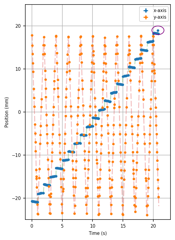
Figure 1: Measured beam positions by the CMOS detector, highlighting the intentionally misdelivered spot.

The CMOS detector was then irradiated with two clinical style fields to assess clinical practicability.

A comparison of the first energy layer between the TPS dose map and the CMOS detector can be seen in Figure 2.
Due to current dead time limitations, a thorough qualitative gamma comparison is not possible owing to missed spots, however many of the key features of the clinical plan (local hot-spots, overall shape) are visible in the CMOS measurement.


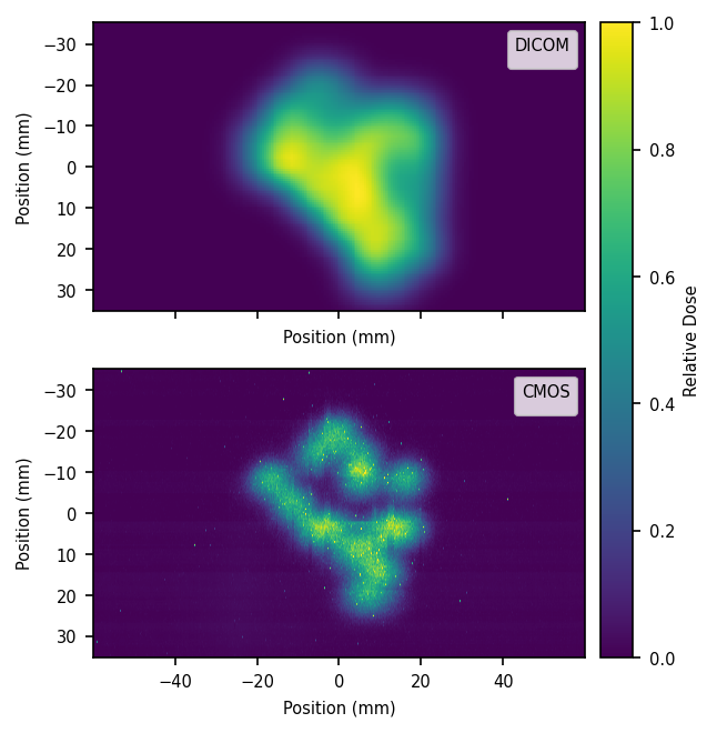
Figure 2: Comparison of measured (CMOS) and expected (TPS) dose distribution.

Conclusion

The vM2428 "LASSENA" CMOS detector demonstrates the viability of the technology for on-line verfication of clinical PBS proton systems.

Development is ongoing to further reduce the dead time between frames.