Vienna, Austria

ESTRO 2023

Session Item

Quality assurance and auditing
6030
Poster (Digital)
Physics
Evaluation of materials suitable for anthropomorphic multimodality (CT/MRI) phantoms for RT purposes
Meshal Alzahrani, United Kingdom
PO-1731

Abstract

Evaluation of materials suitable for anthropomorphic multimodality (CT/MRI) phantoms for RT purposes
Authors:

Meshal Alzahrani1, David Broadbent2, Irvin Teh1, Bashar Al-Qaisieh2, Adrian Walker3, Rachel Lamb3, Richard Speight2

1University of Leeds, Department of Biomedical Imagining Science, Leeds, United Kingdom; 2Leeds Teaching Hospitals NHS Trust, Department of Medical Physics and Engineering, Leeds, United Kingdom; 3Leeds Test Objects Ltd., Leeds Test Objects Ltd., York, United Kingdom

Show Affiliations
Purpose or Objective

With the increasing use of magnetic resonance imaging (MRI) in radiotherapy (RT), there is a need for anthropomorphic multimodality phantoms to be used for various tasks including end-to-end tests of MRI-guided RT workflows. This project aims to find materials that mimic human tissue for MRI and computed tomography (CT) imaging and assess their stability over time and after radiation exposure.

Material and Methods

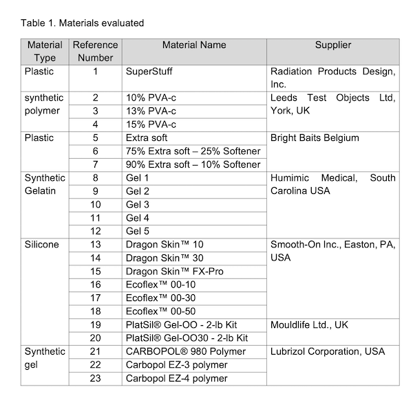
Samples of the materials listed in Table 1 were produced according to the manufacturer's instructions and placed in a test tube. To measure stability over time, the CT number and T1 and T2 relaxation times of the samples have been measured monthly over 7 months. CT scans were acquired using Philips Brilliance BigBore (Philips Medical Systems, Best, Netherlands) with the following settings: 120 kVp, 104 mAs, 2 mm slice thickness, and in-plane resolution of 1.17 mm.

MRI relaxation times were measured using a 3T Siemens Prisma MRI scanner (Siemens Healthineers, Erlangen, Germany). Inversion recovery and Spin-Echo-Multi-Contrast sequences were used to create T1 and T2 maps, respectively. On CT images, T1 and T2 maps, volumes within the materials were drawn, and the mean and standard deviation were measured within these volumes.

To measure stability after radiation exposure, other samples of the same materials were exposed to radiation using an Elekta Versa HD linear accelerator, delivering 10, 100, 250, 500, and 1000 Gy. CT number and MRI relaxation times after radiation exposure were measured.


Results

Only materials 1-4 have CT number, T1 and T2 relaxation times that fell within the range of the brain’s grey and white matter. Figure 1 shows the mean of CT number, T1 and T2 relaxation times, and the standard deviation for each material over time and after radiation exposure.

Regarding the stability of materials over time, all the observed change for all four materials was within two standard deviations (of the pixel values) which indicates the materials are stable over this timeframe. As for the stability after radiation exposure, all the observed changes were within two standard deviations (of the pixel values) except for the T2 relaxation time for material No. 1, and the change that occurred after exposure to 10 Gy in material No. 3. Further investigation will be done to investigate the reasons for these changes.


Conclusion

This project demonstrated the possibility of producing materials that mimic brain tissue in both MRI and CT. Four candidate materials were found; from these, all appear stable over time, and two are stable after radiation exposure.