Accuracy of electron density in a novel high-performance CBCT imaging system
PO-1673
Abstract
Accuracy of electron density in a novel high-performance CBCT imaging system
Authors: Tianyu Zhao1, Alex Price1, Nels Knutson1, Eric Laugeman1, Yao Hao1, Lauren Henke1, Pamela Samson1
1Washington University in St. Louis, Radiation Oncology, St. Louis, USA
Show Affiliations
Hide Affiliations
Purpose or Objective
In this study, we reported the accuracy of the electron density for a novel, high-performance cone beam computed tomography (CBCT) imaging system .
Material and Methods
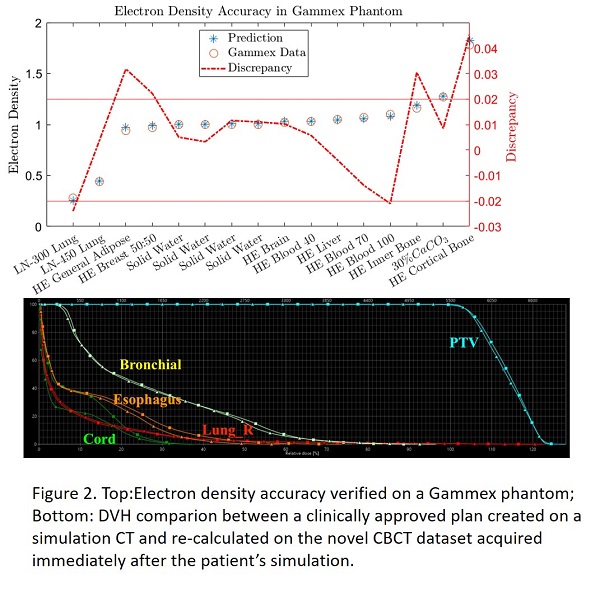
A novel, high-performance CBCT imaging system was designed and installed by the vendor on an o-ring linear accelerator. The imaging panel was reengineered with the size of the panel doubled to 43cm x 86cm. The scintillator layer was changed to CsI of 700 μm in thickness in order to improve the detection sensitivity and imaging resolution. The clinical goal of the new imaging system is to improve image quality to facilitate improved treatment setup with better tumor localization, and to provide a high quality CBCT simulation that can be used for treatment planning/replanning and dose calculation. To verify the accuracy of the electron density of the new CBCT imaging system, Hounsfield units (HU) were measured with a CBCT Electron Density Phantom (SunNuclear, Model 062MA). The phantom is 26.5 cm in the longitudinal direction with optional extension plates to allow adequate scatter in a cone beam geometry. The compositions of the interchangeable inserts were supplied by the vendor. A stoichiometric CT calibration was employed to generate the HU-electron density curve for the two tube voltages commissioned for simulation. Verification was performed by comparing the electron densities measured on a Gammex phantom to the values provided by the vendor. Solid water plates were added to both ends of the Gammex phantom to allow adequate scattering. The measurements were repeated for various clinical protocols with exposures from 50 mAs to 800 mAs. A plan created on a study patient’s helical simulation CT was compared to its verification plan recalculated on the new volumetric CBCT dataset acquired immediately after the simulation in an IRB-approved imaging study.
Results
Figure 1 shows that the variations of the CBCT calibrations are within 0.5% and 0.3% for tube voltage 125kVP and 140kVp, respectively. The difference of the averaged CBCT calibration curves was less than 2% in the bony area between the two tube voltages. The top of the Figure 2 shows the mean discrepancy of the electron densities was 0.0045 with a range between -0.02 and 0.04. The bottom of the Figure 2 shows the DVH comparison between a clinically approved plan created on a simulation CT and re-calculated on a volumetric CBCT dataset from the new imaging system acquired immediately after the patient’s simulation. 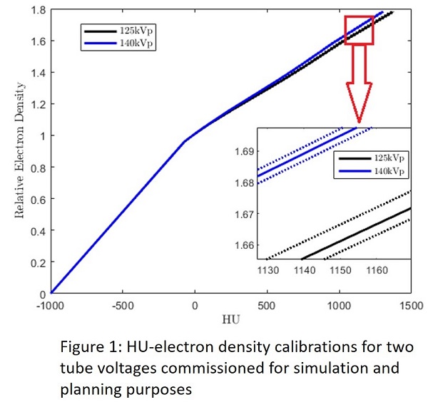
Conclusion
The electron density and resulting dose calculation from the novel CBCT imaging system generated clinically acceptable results.