A pre-processing pipeline for 0.35T pelvic MR image quality enhancement: development and evaluation
PO-1672
Abstract
A pre-processing pipeline for 0.35T pelvic MR image quality enhancement: development and evaluation
Authors: Marica Vagni1, Houng Elena Tran1, Luca Boldrini1, Giuditta Chiloiro1, Andrea D'Aviero2, Alessia Re2, Angela Romano1, Luca Indovina1, Vincenzo Valentini1, Davide Cusumano1, Lorenzo Placidi1
1Fondazione Policlinico Universitario "A. Gemelli" IRCCS, Department of Radiation Oncology, Rome, Italy; 2Mater Olbia Hospital, Department of Radiation Oncology, Olbia (SS), Italy
Show Affiliations
Hide Affiliations
Purpose or Objective
MR-Linac systems offer high soft-tissue contrast volumes thanks to the onboard MR-scanner. However, low frequency intensity inhomogeneity artefacts can occur, resulting in signal losses that may affect the image quality (IQ). Various correction strategies have been proposed to reduce such artefacts on high-field MRIs and very limited cases are focused on 0.35T ones. This study aims to outline and assess an image processing pipeline able to enhance the IQ, offering a better visualization of targets and organs at risk (OARs) and a more reliable intensity distribution for the subsequent digital processing, such as auto-segmentation.
Material and Methods
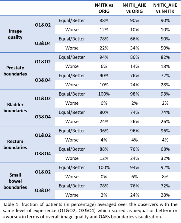
0.35T simulation MRIs (ORIG) from 73 prostate cancer patients treated with MR-Linac were collected. The N4 bias field correction (N4ITK), alone or in conjunction with an Adaptive Histogram Equalization filter (N4ITK_AHE), was applied to the images. Four radiation oncologists with different experience (O1&O2: more than 5 years of experience; O3&O4: less than 5 years) blindly scored 25 randomly selected patients in terms of overall IQ and OARs’ boundaries visibility of the ORIG, N4ITK and N4ITK_AHE images, using a 4-point Visual Grading Analysis scale for the IQ, and a 5-point scale for the OARs. The examined OARs were prostate, bladder, rectum and small bowel. The IQ improvement was also quantitatively evaluated by computing the ratio between the pixels’ intensities standard deviation (σ_ratio) post- and pre-filters application, and the ratio between the Shannon Entropy (SE_ratio) post- and pre-filters application, inside the body.
Results
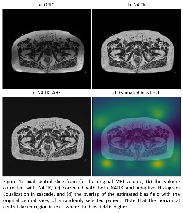
Figure 1 illustrates the effect of filters application on an original image. As shown in Table 1, the application of the N4ITK and N4ITK_AHE with respect to the ORIG resulted mostly in either an improvement or no change of the IQ and the OARs visualization, for all the observers. However, the group O1&O2 scored overall higher as “‘no change/better” compared to the group with less experience. As regards the application of the N4ITK_AHE with respect to the N4ITK, the majority of the scores reflects again no changes or improvements in the rating. From a quantitative perspective, σ_ratio and SE_ratio were both less or equal to 1 for all the 73 patients, with a mean value of 0.72 and 0.93 in the N4ITK/ORIG comparison, respectively, and of 1 in the N4ITK_AHE/N4ITK comparison for both the metrics.
Conclusion
We implemented an image preprocessing pipeline with the aim of facilitating the learning of an automatic segmentation tool without compromising the image visualization for the clinicians. Based on this preliminary work, the N4ITK and N4ITK_AHE application on 0.35T MRIs results mainly in either an improvement or no change of both overall IQ and OARs visualization in clinical practice. Results are also supported by quantitative metrics, showing a decrease in signal dispersion. Future work will investigate the effect of applying this pipeline on the performance of a dedicated AI-based auto-segmentation algorithm.