Long axial field-of-view PET/CT-scanner for deep inspiration breath hold radiation therapy planning
PO-1667
Abstract
Long axial field-of-view PET/CT-scanner for deep inspiration breath hold radiation therapy planning
Authors: Lena Specht1,2, Flemming L. Andersen3,4, Peter A. Andreasen1, Danijela Dejanovic4, Anne K. Berthelsen1, Jeppe Friborg1,2, Mathias G. Askløf4, Peter M. Petersen1, Elisabeth Albrecht-Beste4, Vibeke N. Hansen1, Ivan R. Vogelius1,2, B. Malene Fischer3,4
1Rigshospitalet, Dept. of Oncology, Copenhagen, Denmark; 2University of Copenhagen, Dept. of Health and Medical Sciences, Copenhagen, Denmark; 3University of Copenhagen, Depth. of Health and Medical Sciences, Copenhagen, Denmark; 4Rigshospitalet, Dept. of Clinical Physiology and Nuclear Medicine, Copenhagen, Denmark
Show Affiliations
Hide Affiliations
Purpose or Objective
Objective: Modern, limited and highly conformal radiation therapy (RT) in deep inspiration breath hold (DIBH) has dramatically reduced toxicity for patients with mediastinal lymphomas. However, to enable optimal imaging with fusion of pre- and post-chemotherapy images for RT planning, the pre-chemotherapy staging PET/CT-scan must also be done in DIBH. Ideally, the entire PET-scan would be acquired in breath hold without fusion between sections, but this has not previously been possible. We report here for the first time the advantages of using a long axial field-of-view (LAFOV) PET/CT-scanner for whole body PET/CT-scans in DIBH specifically for treatment planning in DIBH.
Material and Methods
Material & Methods: Patients with mediastinal lymphoma, who may need radiotherapy after their systemic treatment, have their initial staging (pre-chemotherapy) PET/CT performed in DIBH on a flat table-top and in a position suited for later RT. Until now this was performed on a conventional PET/CT-scanner with a field of view of 25 cm. In September 2021, a LAFOV PET/CT (Siemens Biograph Vision Quadra) was installed at our institution. The extended axial coverage (106 cm) and improved sensitivity prompted us to shift the pre-chemotherapy DIBH PET/CT-scans to the new scanner. Our standard procedure is now to perform a whole body DIBH diagnostic CT with i.v. contrast and 4 PET acquisitions in DIBH (each 18 seconds). Here we report on the first clinical experiences with this updated procedure.
Results
Results: The first three patients are presented. Implementation of DIBH on a LAFOV PET/CT scanner is feasible and well tolerated. DIBH PET/CT images of diagnostic clinical quality can be obtained in just 4 x 18 seconds. Clear advantages of the LAFOV DIBH PET/CT scans are:
1. The pre-chemotherapy PET/CT-scan is simpler and faster.
2. Fusion of the pre-chemotherapy PET/CT-scan with the post-chemotherapy DIBH planning CT-scan is simpler and more precise, as the whole pre-chemotherapy scan is made in DIBH. This enables greater accuracy in the contouring of the target for RT
3. The i.v. contrast is visible in all parts of the pre-chemotherapy PET/CT-scan improving the diagnostic quality of the CT-scan, in particular regarding the PET-negative nodes often found in lymphomas
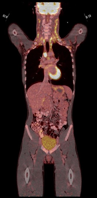
Figure: Long axial field-of-view PET/CT-scan in deep inspiration breath hold of a patient with mediastinal and bilateral neck involvement
Conclusion
Conclusion: Pre-chemotherapy DIBH PET/CT performed on a LAFOV PET/CT scanner makes the post-chemotherapy planning of DIBH RT easier and more accurate, enabling greater conformity and normal tissue sparing.