Development of virtual CBCT simulator and deep-learning-based elemental material decomposition.
PO-1697
Abstract
Development of virtual CBCT simulator and deep-learning-based elemental material decomposition.
Authors: Taisei Shimomura1, Daiyu Fujiwara1, Yuki Inoue1, Atsushi Takeya1, Takeshi Ohta2, Yuki Nozawa2, Toshikazu Imae2, Kanabu Nawa2, Keiichi Nakagawa2, Akihiro Haga1
1Tokushima University, Medical Informatics, Tokushima, Japan; 2The University of Tokyo Hospital, Radiology, Tokyo, Japan
Show Affiliations
Hide Affiliations
Purpose or Objective
Nowadays, Cone-Beam Computed Tomography (CBCT) system plays an important role in image-guided radiation therapy and adaptive radiation therapy (ART). However, the image quality degradation made by the scattered x-ray provides the difficulty in the quantitative dose evaluation. For the quantitative CBCT imaging, in this study, we newly propose the development of a virtual CBCT simulator including the scattered x-ray and show the usefulness in an elemental material decomposition (EMD) by developing a head-and-neck (HN) human phantom library.
Material and Methods
A HN human phantom library including 36 phantoms composing of six elements (H, C, N, O, P, and Ca) was developed by extending the International Commission on Radiological Protection (ICRP) 110 adult female/male phantoms with the information of age, height, and weight in the human population. A CBCT database using this library was then created by a virtual CBCT simulator, which simulates the direct and scattered x-ray on a flat panel detector (FPD) from a raytracing model and a deep-learning (DL) model, respectively. The Gaussian distributed noise on FPD, of which magnitude was evaluated with a cylindrical water phantom of 25 cm diameter using a real CBCT system (Elekta X-ray Volumetric Imaging system), was also included. The usefulness in the virtual CBCT system was demonstrated by applying the developed DL-based U-net EMD model in both virtual phantom and experimental Rando phantom. The evaluation of the EMD results were performed by using the root mean squared error (RMSE) and the structural similarity index measure (SSIM) in each predicted 300 slices in 6 test phantoms.
Results
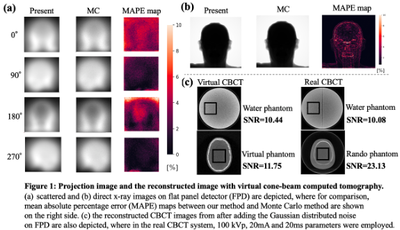
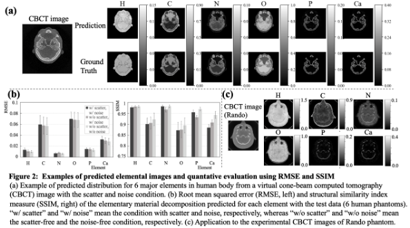
Examples of scattered, direct x-ray and CBCT images with noise produced by the CBCT simulator are shown in Fig.1, where the projection image agreed well with that generated in Monte Carlo (MC) simulation (Fig.1(a), (b)). The noise determined in the water phantom was somewhat large in the CBCT image domain for human phantoms (Fig.1(c)). The elemental map predicted in the DL-based EMD model developed with the virtual CBCT database is shown in Fig.2, where RMSE is less than 0.08 in all elements within the database as shown in Fig.2(a). Although the CBCT image degradation due to the scattered x-ray and the statistical noise slightly affects the prediction accuracy (p<.001 for all metrics except SSIM of N), the amount of these effects was small. The predicted elemental map with the real CBCT image was also generated reasonably well as shown in Fig.2(c).
Conclusion
We demonstrated that the developed virtual simulator can generate various CBCT images based on the human phantom library. Comprehensively, the prediction of the EMD can be successfully performed by preparing the CBCT database from the proposed virtual system, even for the real images using Rando phantom. This study suggests that our approach, to use the computer vision for the data preparation, has a potential in further development of radiation therapy, such as the ART with the EMD from CBCT images.