MRI derived brain morphometric measurements: effect of voxel size, anisotropy and slice orientation
Tamara Lusa Aguero,
Spain
PO-1694
Abstract
MRI derived brain morphometric measurements: effect of voxel size, anisotropy and slice orientation
Authors: Tamara Lusa Aguero1, Steren Chabert2, Alejandro Veloz2
1Hospital Clínico Universitario de Valencia, Radiofísica y Protección Radiológica, Valencia, Spain; 2Universidad de Valparaíso, CINGS, Centro de Investigación en Ingeniería para la Salud, Valparaíso, Chile
Show Affiliations
Hide Affiliations
Purpose or Objective
There is a growing interest in obtaining quantitative indicators of brain volumes in the follow-up of patients undergoing radiation therapy of central nervous system neoplasms, that frequently exhibit radiation-induced atrophy in brain structures. MRI-imaging is the modality of choice.
Acquisition recommendations for volumetric studies are isotropic voxel 1x1x1mm3 and sagittal slice orientation as indicated by the MPRAGE protocol. However, in clinical practice, modifications have been observed due to time constraints, leading to bigger and anisotropic voxels.
Thus, we studied the impact of modifying the acquisition parameters: bigger voxel sizes, slice orientation (axial, sagittal) and anisotropy in volumetric MRI quantifications in different brain structures.
Material and Methods
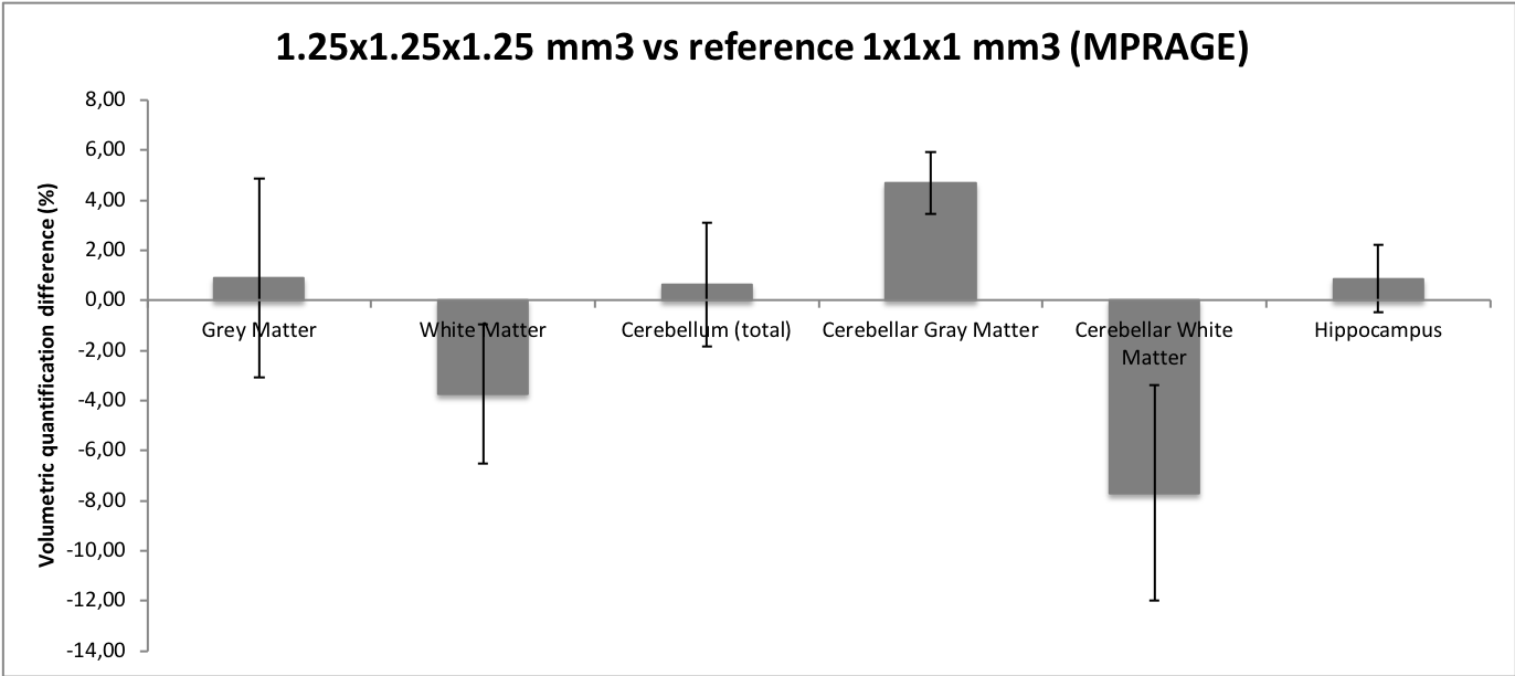
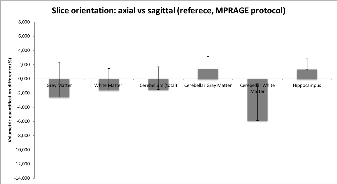
10 healthy subjects underwent 3D-T1-weighted MRI acquisitions. Variations from the MPRAGE protocol were considered, testing bigger voxels (1.1x1.1x1.1 mm3 and 1.25x1.25x1.25 mm3), anisotropic voxels (0.9x0.9x1.2 mm3 and 1x1x1x1.25 mm3) and slice orientation (axial vs sagittal). Volumes were segmented with an automatic software and normalized to the intracranial volume.
Differences in volume estimations were calculated as 100·(Vvariant - Vreference)/Vreference, taking as the reference the recommendations of the MPRAGE protocol: isotropic voxel 1x1x1mm3 and sagittal orientation.
Hotelling statistical significance test was applied.
Results
The cerebellar white-matter was the structure with the greatest range of variation in volumetric quantification when modifying acquisition parameters, observing in average differences of 5.88% ± 2.4 % when performing axial vs sagittal acquisition; 7.7 ± 4.3 % volume underestimation when acquiring with 1.25x1.25x1.25mm3 voxel vs recommended 1x1x1mm3 and 0.6 ± 4.9 % when introducing anisotropy. Other structures, like the gray and white matter of the brain and the hippocampus showed maximum quantification deviations of 4%.
Hotelling test showed no statistically significant differences, but the variations observed are not negligible.
Conclusion
The use of isotropic and smaller voxels may be time consuming, but the inaccuracies introduced in quantitative volume estimations due to changes in acquisition parameters must be kept in mind, as they may be greater than the volumetric reduction due to radiation-induced atrophy, which is subject of study, and may lead to a misdiagnosis, resulting highly advisable not to vary the acquisition parameters from those recommended by the literature.