Characterization of A Novel High-Performance Cone Beam Computed Tomography Imaging System
PO-1684
Abstract
Characterization of A Novel High-Performance Cone Beam Computed Tomography Imaging System
Authors: Tianyu Zhao1, Alex Price1, Eric Laugerman1, Yao Hao1, Nels Knutson1, Hua Li1, Lauren Henke1, Pamela Samson1
1Washington University in St. Louis, Radiation Oncology, St. Louis, USA
Show Affiliations
Hide Affiliations
Purpose or Objective
Image quality of a novel, high-performance cone beam computed tomography (CBCT) imaging system was measured and reported in this study.
Material and Methods
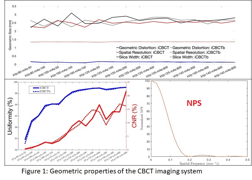
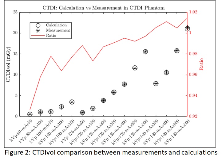
A re-engineered CBCT imaging panel with the doubled to 86cm x43cm for a full field of view (FOV) of 55.8cm in diameter and extendable to 70 cm was installed by the vendor on a commercial o-ring linear accelerator. A thicker layer of CsI crystal scintillator replaced Gadolinium Oxysulfide for better light yield and higher imaging resolution. The gantry rotation speed was increased to 10 rotations per minute. Combined with the new imaging panel, a 211o rotation was deemed enough for a full CBCT reconstruction. This improvement reduced the acquisition time to 5.9 seconds. A new reconstruction mode based on a linear Boltzmann transport solver was introduced to reduce the imaging artifact from scattering. The CBCT system provides four tube voltages ( 80 kVp, 100 kVp, 125 kVp and 140 kVp) for image-guided radiotherapy (IGRT) and the latter two of the four tube voltages was used for planning (CBCTp) purpose. In this study, we tested the important imaging quality parameters such as imaging distortion, resolution, slice thickness, uniformity, and the contrast-to-noise ratio reconstructed with iterative CBCT (iCBCT) and iterative CBCT on a Boltzmann transport equation solver (iCBCTAcuros) reconstructions on Catphan modules 528, 404, and 486. Tube voltages, exposures, scanning speed, and imaging dose were measured in direct comparison to the parameters provided by the vendor. Noise levels were measured with the same Catphan, and noise power spectrum (NPS) was calculated on a cylindric water phantom scaned with all clinical protocols.
Results
Imaging distortion and slice thickness were measured less than 0.2 mm and 2.5mm in all imaging protocols with both reconstruction modes. Imaging resolution was measured 1.35mm with iCBCTAcuros and 2.5mm with iCBCT mode. The maximum deviation in the measured Tube voltage and exposure was 1.7 kVp and 2.3 mAs, respectively. The scanning speed was measured consistently between 5.85 s and 5.86s with all clinical protocols. The congruency of imaging dose was within 2%, except for tube voltage of 80kVp, where the measured average imaging dose differed 6% from the vendor’s calculation. Uniformity was measured above 98%, between 97% and 98%, and between 95% and 97% in imaging protocols with tube voltage of 125kVp and above, 100 kVp, and 80 kVp. The contrast-to-noise ratio was 0.5 at 80 kVp and 50 mAs, but increased rapidly with tube voltage and exposure. NPS analysis showed that the imaging noise is dominated by white noise with a very weak peak at 0.25mm-1 in a magnitude of 1.8% of the white noise, indicating very weak imaging artifacts compared to current CBCT images.
Conclusion
The novel CBCT system met expected image quality performance and is expected to achieve acquisition in a single-breath hold. The imaging dose is 2~3 fold lower than the currently installed CBCT systems on a C-arm Linacs.