MRI for 3D target temperature assessment in hyperthermia of locally advanced cervical cancer
Carolina Carrapiço-Seabra,
The Netherlands
PO-1681
Abstract
MRI for 3D target temperature assessment in hyperthermia of locally advanced cervical cancer
Authors: Carolina Carrapiço-Seabra1, Iva VilasBoas-Ribeiro1, Anton Rink1, Martine Franckena1, Sergio Curto1, Gerard C. van Rhoon1,2
1Erasmus MC Cancer Institute, University Medical Center Rotterdam, Department of Radiotherapy, Rotterdam, The Netherlands; 2Faculty of Applied Sciences, Delft University of Technology, Department of Radiation Science and Technology, Delft, The Netherlands
Show Affiliations
Hide Affiliations
Purpose or Objective
Hyperthermia, the elevation of tumour temperature up to 43ºC, is used as a sensitising cancer treatment that enhances the effects of radiotherapy and chemotherapy. Clinical trials have shown the correlation of achieved temperatures and thermal dose with treatment outcome. In the treatment of locally advanced cervical cancer, temperature monitoring is performed using thermal probes placed in intraluminal locations. Although these intraluminal measurements have been correlated with tumour temperatures, thermal probes provide limited information since only few point locations can be measured. Currently, the use of Magnetic Resonance (MR) imaging is the only clinical option to measure 3D temperatures non-invasively. The aim of this study was to (1) validate the correlation between intraluminal probes and MR-based temperatures in the target and (2) investigate the prediction role of MR based temperature metrics for thermal dosimetry.
Material and Methods
This study included seven patients diagnosed with locally advanced cervical cancer that were treated using hyperthermia as sensitiser to radiotherapy. The hyperthermia treatment was performed in the MR‑compatible BSD‑2000‑3D hyperthermia system integrated into a 1.5T GE Signa Excite scanner. During the hyperthermia treatment, two temperature monitoring techniques were used: intraluminal and MR- thermometry. Intraluminal thermometry was acquired using Bowman probes inserted into closed tip catheters placed in the vagina before the hyperthermia treatment. MR thermometry was obtained following the proton resonance frequency shift method. The target temperature (Hyperthermia Target Volume: HTV) was obtained from the MR thermometry and its correlation with the temperature measured in the vaginal probe was calculated. The temperature achieved in 50% of the target volume (T50) was also calculated to investigate the efficacy of treatment.
Results
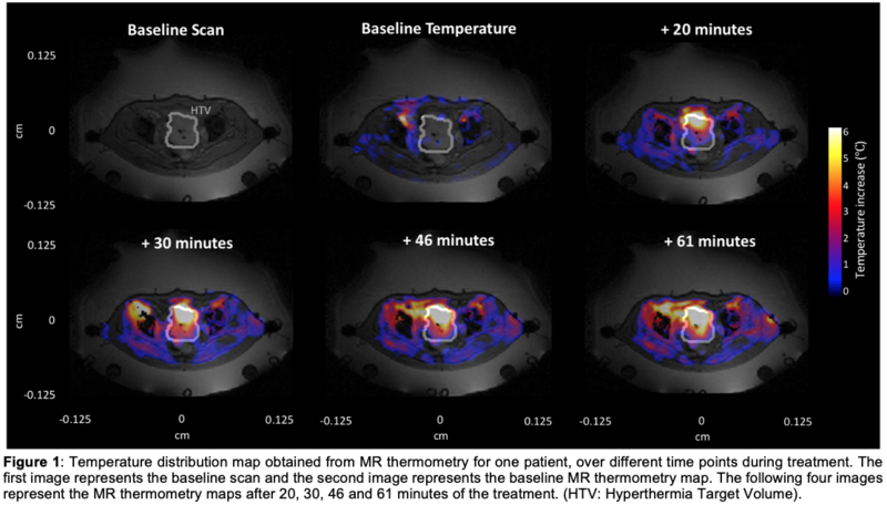
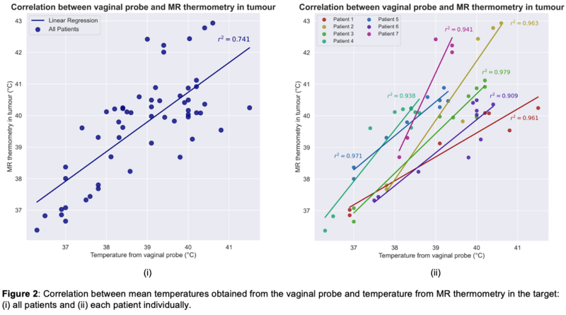
Figure 1 shows the temperature distribution map obtained from MR thermometry for one representative patient, over different time points during treatment. The correlation between mean temperatures obtained from the vaginal probe and MR thermometry in the HTV are shown in Figure 2. When all the seven patients were evaluated, a correlation coefficient of 0.74 was obtained. Nevertheless, when evaluating the correlation for each patient individually, the coefficient values ranged between 0.91 and 0.98. These greater values are explained by differences in patient condition and characteristics such as treatment tolerance and tumor physiology (e.g., perfusion and size) and patients’ thermoregulation. The T50 obtained from the target was 40.5ºC, (compared to 39.6 ºC from the vaginal probe) which indicates a good target coverage.
Conclusion
This study showed that MR thermometry in the target region is comparable to vaginal intraluminal thermometry. Therefore, MR thermometry facilitates the 3D target temperature assessment, enabling thermal dosimetry and improving treatment efficacy.