Quantitative analysis of vaginal and uterine geometry for patient-tailored BT applicator development
Sharline van Vliet-Perez,
The Netherlands
PO-2150
Abstract
Quantitative analysis of vaginal and uterine geometry for patient-tailored BT applicator development
Authors: Sharline van Vliet - Perez1,2, Tim Crone1, Ruben van den Broek1, Robin Straathof1,2, Linda Wauben2, Nick van de Berg2, Jenny Dankelman2, Ben Heijmen1, Inger-Karine Kolkman - Deurloo1, Remi Nout1
1Erasmus MC Cancer Institute, University Medical Centre Rotterdam, Radiotherapy, Rotterdam, The Netherlands; 2Delft University of Technology, BioMechanical Engineering, Delft, The Netherlands
Show Affiliations
Hide Affiliations
Purpose or Objective
The currently used applicators for locally advanced cervical cancer (LACC) brachytherapy (BT) do not optimally align with individual patient anatomy and have fixed catheter positions and angles limiting treatment plan conformity. Patient-tailored applicators with optimally planned catheter channels could be the solution for this problem. To develop patient-tailored applicators, and to create a representative data set for safety tests and quality management of the patient-tailored applicators, data on the dimensions of the distended vaginal and uterine shape is required. In previous studies, magnetic resonance imaging (MRI) has been successfully used to acquire data on the shape and dimensions of the vaginal cavity and uterus in a healthy population. However, little is known about the distended geometry of LACC patients after concomitant external beam radiation therapy and chemotherapy. Therefore, the aim of this study is to evaluate the vaginal and uterine geometry distended with ultrasound gel in LACC patients.
Material and Methods
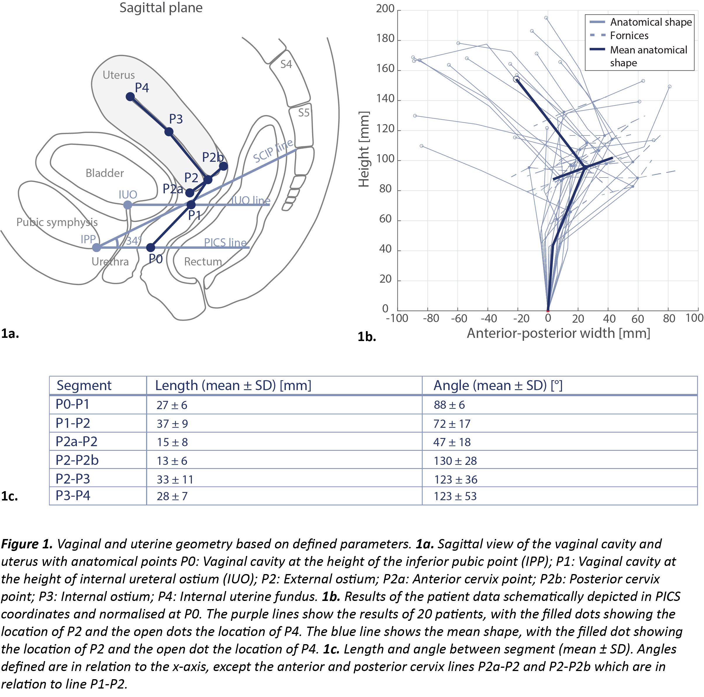
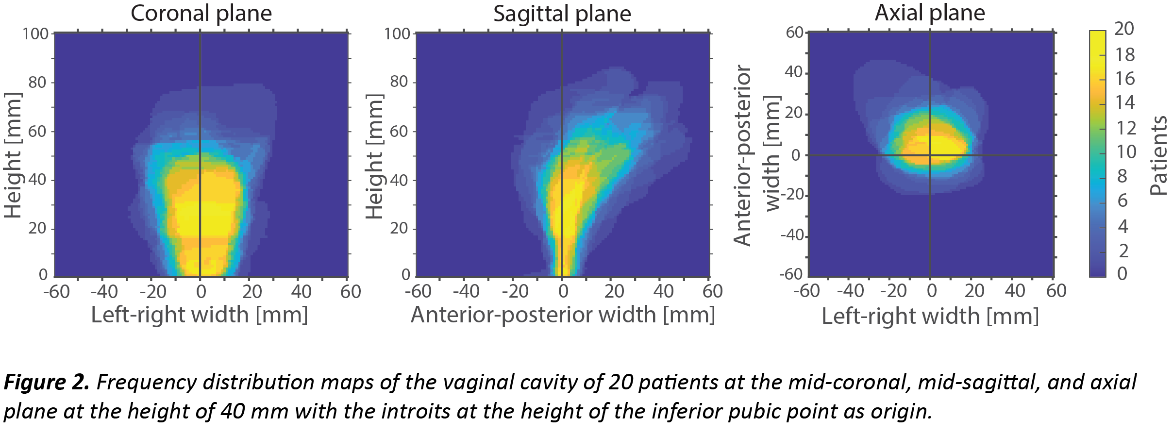
In total 20 pre-BT MRIs of LACC patients with ultrasound gel inserted in the vaginal cavity were included. The pre-BT MRIs were taken approximately one week before BT treatment. For analysis, the orientation of the MRIs was corrected based on the Pelvic Inclination Correction System (PICS). A set of parameters describing the vaginal and uterine geometry was selected (Figure 1a). The lengths and angles of mid vagina (P0-P1), upper vagina (P1-P2), anterior cervix (P2a-P2), posterior cervix (P2-P2b), endocervical canal (P2-P3), and uterine body (P3-P4) were determined. Furthermore, the left-right (LR) width at point P0, P1, and P2 were evaluated. In addition, frequency distribution maps of the vaginal cavity were made based on 3D models of the delineated MRIs.
Results
In Figure 1, the lengths and angles between the vaginal and uterine segments are depicted. The LR width (mean ± SD) was 22 ± 7 mm, 35 ± 8 mm, and 34 ± 16 mm at P0, P1, and P2 respectively. In Figure 2, frequency distribution maps of the 3D models of the vaginal cavities are depicted. The mean volume of the 3D models was 48 (range: 12-69) cm³.
Conclusion
In this study the distended vaginal and uterine geometry was evaluated in LACC patients. The large inter-patient variation in length, angle, and width of different sections of the vaginal cavity suggests a patient-tailored design, especially in the top part of the vaginal cavity. The largest variation was seen in the region P2-P4 as indicated by the length of P2-P3, the width at P2, the angle in P3-P4, and the frequency distribution maps. The evaluated parameters and frequency distribution maps are now available and can be used as design parameters and for safety and quality assessment of the patient-tailored applicators for LACC BT.