Intra-fraction variation of volume and position of stomach, gastric gas and gastric contents on MRI
Joanna Kucharczak,
United Kingdom
PO-1899
Abstract
Intra-fraction variation of volume and position of stomach, gastric gas and gastric contents on MRI
Authors: Joanna Kucharczak1, Cynthia Eccles2, Ananya Choudhury2, Mairead Daly3
1University of Cambridge, School of Clinical Medicine, Cambridge, United Kingdom; 2The University of Manchester, Division of Clinical Cancer Sciences, Manchester, United Kingdom; 3The University of Manchester, Division of Clinical Cancer Sciences , Manchester, United Kingdom
Show Affiliations
Hide Affiliations
Purpose or Objective
Size and position inter-fraction variation of abdominal organs at risk (OARs), including the stomach have been reported (1, 2), causing variation in dose received (3). Limited data exist on intra-fraction variation. This work quantifies and compares intra-fraction position and volume variation of stomach, gastric gas, and gastric contents on MRI with an abdominal compression belt.
Material and Methods
Four participants, 3 patients with hepatocellular carcinoma and one healthy volunteer, were imaged on the Unity MR-Linac (Elekta Limited, Crawley, UK) under an institutional ethically approved study (NCT04748094). Images were acquired at 0, 20 and 30 minutes after set up on each visit. An abdominal compression belt was used following local protocols for all image timepoints, and patients were asked to refrain from eating or drinking for a minimum of 2 hours prior. Three participants attended for 2 visits and one attended for one visit. The stomach was delineated on 3D T2-weighted MRI scans by a single observer. Structures were checked and amended by a second observer 8 weeks later. Gastric gas and contents were delineated using signal intensity value thresholding (<60 for gas and <175 for contents). The volumes and centre of mass (COM) positions for stomach, gastric gas and gastric contents were recorded for each patient at each timepoint for all imaging sessions. The mean intra-fraction volume motion and COM position from the 0 minute timepoint and the average percentage changes of intra-fraction volume between different timepoints were calculated.
Results
The population mean variations in stomach volume from the 0 minute timepoint were 3.16ml (range: 0.33-9.49), 3.15ml (0.60-6.31), and 3.54ml (0.03-5.79) for intra-fraction stomach, gastric gas, and gastric contents volumes respectively.
The averages of the mean magnitude of movement from the 0min timepoint were 0.21cm (range: 0-0.31), 0.41cm (0.21-0.72), and 0.30cm (0.23-0.39) for intra-fraction stomach, gastric gas, and gastric contents COM positions respectively. Minimal baseline drift of stomach COM in the craniocaudal direction was seen.
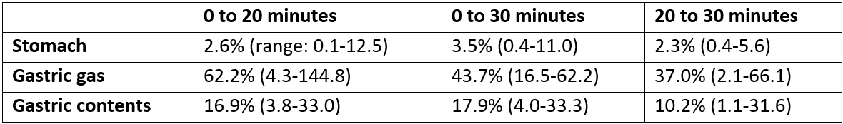
The volume changes are shown in Table 1.
Table 1: Average percentage changes in stomach, gastric gas and gastric contents volumes for 0 to 20 minutes, 0 to 30 minutes and 20 to 30 minutes timepoints.
Conclusion
Stomach position and volume are stable throughout a 30 minute duration in fasted patients imaged with abdominal compression. Some variation is observed in the intra-fraction gastric gas and gastric contents volume. Further studies are required to assess the difference in dosimetry at each timepoint with respect to the volume and COM position changes. The minimal intra-fraction stomach variation observed in this study is unlikely to affect the dose received to the tumour and the OARs.