Surface-guided imaging with ExacTrac Dynamic System
PO-1892
Abstract
Surface-guided imaging with ExacTrac Dynamic System
Authors: Eun Young Han1, Dong Joo Rhee1, Shane Krafft1, Tina Briere1
1The University of Texas MD Anderson Cancer Center, Radiation Physics, Houston, USA
Show Affiliations
Hide Affiliations
Purpose or Objective
ExacTrac Dynamic is an in‐room X‐ray and surface/thermal imaging device that offers X‐ray and surface‐based positioning and monitoring. The purpose of this study was to verify surface tracking and X‐ray imaging accuracy through an end-to-end test following intentional motions for two treatment workflows. The accuracy of surface tracking on phantoms with various skin tone and textures was also studied.
Material and Methods
A 2-target stereotactic brain treatment plan was delivered with Standard X-ray workflow and CBCT positioning with X-ray monitoring workflow. The plan was generated for an RTsafe head phantom, and an ionization chamber was placed at the center of the superior target with a 1.3 cm radius. The phantom was placed on a motorized motion platform to create four discrete intra-fractional motions while beam was on. Both measurements were compared to a measurement without ExacTrac and intra-fractional motion (reference).
To verify the accuracy of surface tracking for various skin stones (bright/dark), textures (dry/moist) and camera temperature equilibrium states (state B [camera on <10min] and state C [>1hr]), we measured the difference between surface tracking and X-ray image registration for bright- and dark-skinned phantoms. The bright/dark-skinned phantom was simulated by applying a makeup foundation to the surface of the RTsafe head phantom (Figure 1). After stereoscopic x-ray imaging reset the surface tracking to the planned position, the phantom position is changed and the discrepancy of surface tracking against X-ray imaging were recorded. The measurements were repeated for couch angles of 0°, 45°, and 270°.
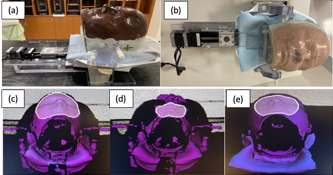
Figure 1 Motion platform with dark(a) and bright-skinned phantom(b), surface imaging with bright(c), dark and moist(d) and dark and dry-skinned phantom(e).
Results
Measured doses were 2078 cGy for X-ray workflow and 2053 cGy for CBCT workflow. Both measured doses were within 1.4% of the reference measured dose (2082 cGy). We observed that the tracking accuracy was compromised due to surface degradation and region-of-interest flickering for the dark moist-skinned phantom with extended time (camera state C). This caused fluctuations in surface tracking and false beam interlocks (Figure 1).
The difference between X-ray and surface imaging was about 0.17±0.11mm/0.03±0.05° for the bright-skinned phantom, whereas those for the dark dry-skinned phantom were 0.26±0.14mm/0.05±0.09° and for the dark moist-skinned phantom 0.53±0.21mm/0.07±0.08°. The differences were larger for couch angles of 0° and 45° in the lateral and longitudinal directions (Table 1).
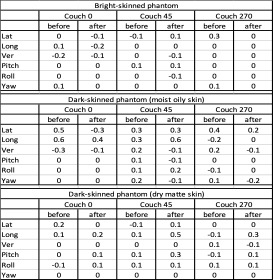
Table 1 Difference in surface tracking compared to X-ray image verification before and after intentional shifts.
Conclusion
The results demonstrated that surface tracking could differ by up to 0.5 mm depending on skin tone, texture, and camera equilibrium state. Thermal imaging of real patients may reduce this difference. X-ray image verification should be rigorously used with surface tracking for dark moist-skinned patients.