The right hemidiaphragm top is an accurate tracking surrogate for abdominal organ motion
Irma van Dijk,
The Netherlands
PO-1874
Abstract
The right hemidiaphragm top is an accurate tracking surrogate for abdominal organ motion
Authors: Jeffrey Veldman1, Zdenko van Kesteren1, Ebony Gunwhy1, Michael Parkes1, Markus Stevens2, Joost van den Aardweg3, Geertjan van Tienhoven1, Arjan Bel1, Irma van Dijk1
1Amsterdam UMC - location University of Amsterdam, Department of Radiation Oncology, Amsterdam, The Netherlands; 2Amsterdam UMC - location University of Amsterdam, Department of Anaesthesiology, Amsterdam, The Netherlands; 3Amsterdam UMC - location University of Amsterdam, Department of Pulmonology, Amsterdam, The Netherlands
Show Affiliations
Hide Affiliations
Purpose or Objective
Radiotherapy of thoracic and upper abdominal tumours is challenging due to respiration-induced tumour motion. Tracking using kilo voltage (kV) imaging can be used to deal with respiratory motion. However, abdominal organs are difficult to visualize using kV imaging. Instead, the diaphragm could serve as surrogate for abdominal organ/tumour motion, provided the error of such a surrogate is known. Particular challenges in establishing such errors during free breathing (FB) can be addressed when using prolonged breath-holding (PBH; 3-12 minutes), where the diaphragm drifts in cranial direction by approximately 3mm/min. The aim of this study was to quantify the error when using the right hemidiaphragm top (RHT) as surrogate for abdominal organ motion during PBH, with the intention to potentially apply PBH in radiotherapy with tracking.
Material and Methods
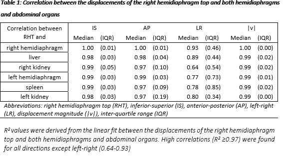
After METC approval and signing informed consent, 15 healthy volunteers were included and trained to perform PBHs during two magnetic resonance imaging (MRI) sessions (PBH-MRI1 and PBH-MRI2). From each session, we selected seven images (dynamics) to determine organ displacement during PBH by using deformable image registration (DIR). On the first dynamic, we segmented the RHT, right and left hemidiaphragm, liver, spleen and both kidneys. We used the deformation vector fields (DVF) generated by DIR to quantify organ displacement between two dynamics in inferior-superior (IS), anterior-posterior (AP) and left-right (LR) direction and calculated the 3D vector magnitude (|v|). We used a linear fit to compare displacements of the RHT and each organ and determined the correlation (R2 of the fit) and the displacement ratio (DR, slope of the fit) between the displacements of the RHT and each organ. We quantified the difference between the DRs of PBH-MRI1 and PBH-MRI2. Additionally, we estimated organ displacement in PBH-MRI2 by applying the DR from PBH-MRI1 to the displacement of the RHT in PBH-MRI2. We then compared the estimated displacement to the measured organ displacement in PBH-MRI2. The difference between these values is defined as the estimation error when using the RHT as surrogate to estimate abdominal organ displacements.
Results
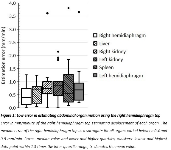
All organs showed a linear drift over time during PBH. We found strong correlations between displacements of RHT and each organ (R2>0.96) in the IS and AP direction and |v|, and strong to moderate correlations in the LR direction (0.93 > R2 > 0.64) (Table 1). The median DR difference between PBH-MRI1 and PBH-MRI2 varied between 0.13-0.31 for all organs. The median estimation error of the RHT as surrogate varied between 0.4-0.8 mm/min for all organs (Figure 1).
Conclusion
The small estimation error established in this study shows that the RHT could serve as an accurate surrogate for abdominal organ motion in radiotherapy using tracking, provided the estimation error is taken into account in treatment margins.