Deformation of co-irradiated lymph nodes in AI-based online adaptive radiotherapy of bladder cancer
Katrine Michella Gullitsø,
Denmark
PO-1873
Abstract
Deformation of co-irradiated lymph nodes in AI-based online adaptive radiotherapy of bladder cancer
Authors: Katrine Michella Gullitsø1, Tine Louise Tofte2, Lina Frida Möller Åström2, Anja Nielsen3
1Herlev & Gentofte Hospital, Department og Oncology, Radiotherapy, Herlev , Denmark; 2Herlev & Gentofte Hospital, Department of Oncology, Radiotherapy , Herlev , Denmark; 3Herlev & Gentofte Hospital , Department of Oncology, Radiotherapy , Herlev , Denmark
Show Affiliations
Hide Affiliations
Purpose or Objective
The purpose of this study was to evaluate automatic deformation of pelvic lymph nodes in AI-based, CBCT-guided online adaptive radiotherapy (oART) of bladder cancer, by comparing deformed target structures with structures delineated by an oncologist. Since the pelvic lymph nodes are expected to be immobile relative to bones, the corresponding target volumes were expected not to change significantly during the course of treatment. However, observations by the physicists and RTT’s motivated a hypothesis that a difference in occurs.
Material and Methods
This retrospective study included 10 consecutive patients with bladder cancer and co-irradiated pelvic lymph nodes, treated from January 2020 to March 2022. Reference clinical target volumes(CTV-ref) with iliac, obturator and pre-sacral nodes were delineated by an oncologist on pre-treatment CT scans. During the daily oART treatments, these target structures were automatically deformed to fit the anatomy of the CBCT, generating adapted versions of the structures (CTV-adp). CTV-ref and CTV-adp were compared using difference in volume, center of mass (COM) shift and dice similarity index (DSI). Corresponding planning target volumes (PTV-ref and PTV-adp) with margins of 8 mm in superior and inferior directions and 3 mm in the anterior, posterior and lateral directions were generated and compared.
Results
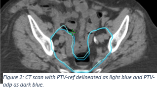
The mean (range) volume for CTV-ref was 385.38 (285.70 - 510.70) cm3. Over all patients, the mean (range) volume difference between CTV-ref and CTV-adp was 5.66 (1.16-19.79) cm3, corresponding to 1.24% (0.29%-5.23%)(Figure 1). Eight patients showed a significant difference in volume (p <0.05) between CTV-adp and CTV-ref. For two of the eight patients, the mean CTV-ref was smaller than the mean CTV-adp. For the remaining 6 patients, the mean CTV-adp was larger than the mean CTV-ref. The COM shift varied widely among the patients and in the different directions. In the lateral direction, the COM shift ranged from -0.22 cm to 0.25 cm, with a mean value of -0.01 cm. In the anterior/posterior direction, it ranged from -0.33 cm to 0.26 cm, with a mean value of -0.05 cm, while in the cranio/caudal direction it ranged from -0.23 cm to 0.24 cm with a mean value of 0.00 cm. The mean (range) DSI was 0.91 (0.86-0.94). The mean (range) volume for PTV-ref was 740.20 (564 - 935) cm3. Comparing PTV-ref and PTV-adp, the mean (range) volume difference was -6.79 (2.4 - -48.14) cm3, corresponding to -0.99% (0.30% - -6.48%) (Figure 2). While PTV-adp in general was smaller than PTV-ref, the mean difference was small.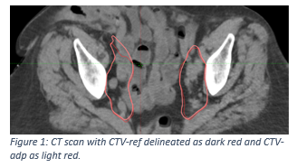
Conclusion
There was a significant difference between CTV-adp and CTV-ref for a majority of the patients. While CTV-adp in general was larger than CTV-ref, PTV-adp was often smaller than PTV-ref. It can thus be concluded that the position of the volume difference in the CTV is crucial for its effect on the PTV and consequently potentially the dose distribution.