Evaluation of a new AI-based sCT generator for MR-only radiotherapy workflows
PO-1828
Abstract
Evaluation of a new AI-based sCT generator for MR-only radiotherapy workflows
Authors: Alban Roussel1, Stephane Dufreneix1, Arthus Cannard2, Thaïs Roque2, Kundar Shreshtha2, Nikos Paragios2,3, Camille Guillerminet1, Damien Autret1
1Institut de Cancérologie de l'Ouest, Medical Physics, Angers, France; 2Therapanacea, Clinical and Partnerships Affairs, Paris, France; 3Centrale Supelec, University of Paris-Saclay, Gif-sur-Yvette, France
Show Affiliations
Hide Affiliations
Purpose or Objective
MR-only radiotherapy workflows require specific MRI sequences to generate a synthetic CT (sCT) on which dose calculation can be performed. To add this sequences, the MR exam time is thus increased up to 15 min and registration is needed between the sCT and the MR sequence on which contours are delineated. The objective of this study is to evaluate a new deep-learning based sCT generator based on MR sequence used for contouring.
Material and Methods
90 patients were enrolled in the study for two locations (40 brain and 50 pelvis, including some hip prostheses). MR sequences used for contouring and sCT generation were T2 Space 3D (pelvis) and T1 Gd 3D (brain). The synthetic-CT AI-model was trained using end-to-end ensembled self-supervised GANs endowed with cycle consistency with planning CTs and T2w-MRIs (pelvis) and T1w-MRIs (brain). For the analysis, sCTs and CTs were rigidly registered and compared. HU mean errors (ME) and mean absolute errors (MAE) between sCTs and CTs were estimated for various structures: body/PTV/brain/skull for brain patients and body/PTV/rectum wall/femoral heads/bladder for pelvis patients. A dosimetric analysis was also performed by reporting DVH metrics Dmean and D98% for PTV and OARs as well as a 2D 1%/1mm gamma analysis with a 2% threshold.
Results
MAE for the body was 36.3 HU for the brain and 16.5 HU for the pelvis. Largest HU ME errors were observed for the bones (skull and femoral heads respectively) (Figure 1).
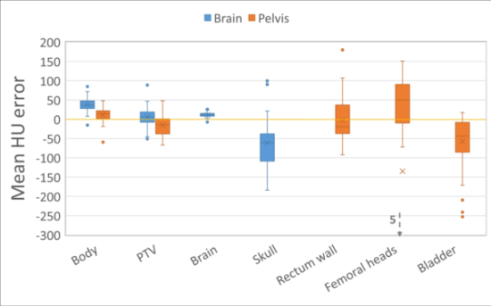
Figure 1 : boxplots of the mean UH errors for various structures of the brain and pelvis cohorts
The Dmean mean error was smaller than 2 % for PTVs and OARs but Dmean deviations could reach up to 15% for femoral heads (Figure 2). The mean D98% difference was -1.4% for the brain PTVs and -3.5% for the pelvis PTVs. The mean gamma passing rate was 90.8% for the brain and 80.2 % for the pelvis.
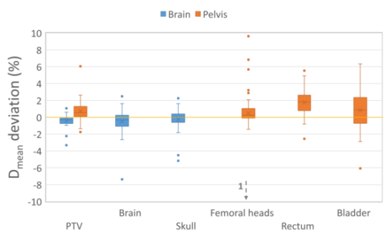
Figure 2 : boxplots of Dmean deviations collected for various various structures of the brain and pelvis cohorts
Conclusion
This study demonstrated the feasibility of using AI-tools to generate clinically acceptable synthetic CT for MR-only radiotherapy workflows in the pelvic and brain regions. Deviations between sCT and CT were comparable to those reported in the literature for both image and dosimetric evaluations. Future work will include the comparison of this new sCT generator to other AI and non-AI solutions, as well as investigate the ability of using sCTs for patient positioning.