Neutron contribution in pediatric proton therapy and field angle dependence
PO-1821
Abstract
Neutron contribution in pediatric proton therapy and field angle dependence
Authors: Johannes Tjelta1,2, Kristian S. Ytre-Hauge2, Franz S. Englbrecht3, Katia Parodi3, Georgios Dedes3, Erlend Lyngholm2, Andreas H. Handeland2,1, Helge Henjum2, Camilla H. Stokkevåg2,1
1Haukeland University Hospital, Department of Oncology and Medical Physics, Bergen, Norway; 2University of Bergen, Department of Physics and Technology, Bergen, Norway; 3Ludwig-Maximilians-University of Munich, Department of Medical Physics, Munich, Germany
Show Affiliations
Hide Affiliations
Purpose or Objective
The out-of-field dose contribution from neutrons is a byproduct of proton therapy, and due to their lack of charge, neutrons have a longer mean free path and can potentially contribute to adverse late effects in organs distant from the target. In addition, the neutron biological effectiveness varies considerably depending on neutron energy. This study aimed to examine the angular dependency for absorbed and equivalent doses on pediatric brain tumor patients treated with intensity-modulated proton therapy.
Material and Methods
Treatment plans optimized in Eclipse (Varian, Palo Alto, CA) were recalculated with the FLUKA Monte Carlo code to obtain neutron absorbed and biologically equivalent dose distributions produced in the patient (2.5mm³ scoring voxels). The planning CT scan included the cranium down through the pelvis, with 54 Gy(RBE) prescribed to a glioma brain tumor. Two target scenarios were delineated and optimized; the original clinical target volume (CTV) (1.8 cc) and a larger CTV (4.4 cc). Three pencil beam scanning plan configurations were simulated for each of the two CTV scenarios: One field either from posterior (0°), 45° or vertex (90°), combined with additionally two lateral fields (80° and 280°). Equivalent dose calculations were done online in FLUKA, where each neutron interaction was applied energy-specific radiological weighting factors (w_R) based on the ICRP 103 report. Single field optimization was used and all neutron doses were calculated as dose to medium.
Results
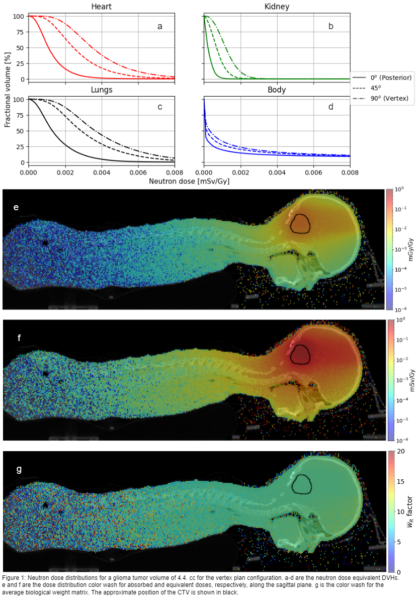
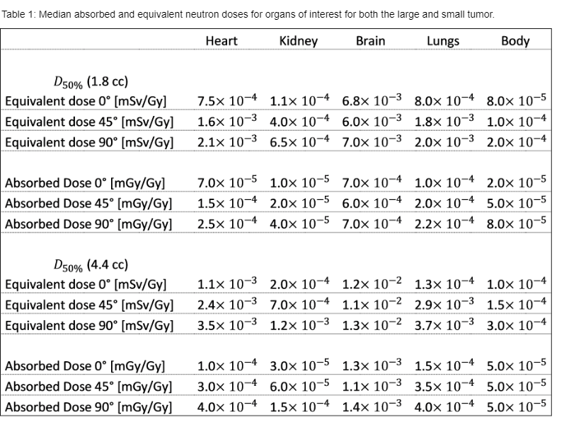
The brain was exposed to the highest neutron dose regardless of field angle, approx. 1.2 μSv/Gy (4.4 cc). Neutron dose distributions varied depending on field configuration (Figure 1a-d). The difference between the posterior and vertex field with equivalent doses at D50% ranged between a factor of 2.5 and 6 depending on organ (Table 1), with the difference increasing further away from the tumor, heart at 3.5 μSv/Gy and kidney at 1.2 μSv/Gy (4.4 cc, 90°). The same trend was observed for absorbed doses but with less difference, ranging from 2 to 5. The median equivalent dose to the heart for the whole treatment of the small tumor was estimated to be 0.06 mSv for the posterior plan and 0.2 mSv for the vertex plan. With respect to tumor, out-of-field doses scaled as a function of the volume irradiated and were 1.5-2.5 times greater for a larger tumor (Table 1). The equivalent doses ranged from 8-11 times higher than the absorbed doses (Figure 1e-g).
Conclusion
The field angle impacted the neutron dose, and the vertex configuration had the most substantial contribution to out-of-field doses. All plan scenarios resulted in total neutron equivalent doses below the dose exposure from an average pediatric CT scan (reference 2-10 mSv). Nevertheless, as consequences of undesired neutron dose deposition and biological effectiveness in healthy tissues remains uncertain, out-of-field neutron contribution should be closely monitored as proton delivery techniques develops.