Dosimetric verification of a Monte Carlo treatment planning system for MR-guided proton therapy
PO-1816
Abstract
Dosimetric verification of a Monte Carlo treatment planning system for MR-guided proton therapy
Authors: Hermann Fuchs1, Fatima Padilla-Cabal1, Brad Oborn2,3,4, Erik Traneus5, Dietmar Georg1
1Medical University of Vienna, Department of Radiation Oncology, Vienna, Austria; 2OncoRay – National Center for Radiation Research in Oncology, Faculty of Medicine and University Hospital Carl Gustav Carus, Technische Universität Dresden, Helmholtz-Zentrum Dresden-Rossendorf, Dresden, Germany; 3University of Wollongong, Centre for Medical Radiation Physics (CMRP), Wollongong, Australia; 4Illawarra Cancer Care Centre, (ICCC), Wollongong, Australia; 5RaySearch Laboratories AB, Research, Stockholm, Sweden
Show Affiliations
Hide Affiliations
Purpose or Objective
The combination of on-bed MR-imaging with proton therapy (MRgPT) may allow the next step in treatment fidelity. In such a conceptual design the magnetic field does not only influence the spatial dose deposition; in addition the path of the primary treatment beam requires special modelling during dose calculation for treatment planning. We report the first results of a dosimetric verification of a Monte Carlo based treatment (MC) planning system (TPS) for MRgPT, i.e. a research version of RayStation (RaySearch Laboratories AB).
Material and Methods
An electromagnet was positioned in front of a horizontal pencil beam scanning (PBS) research beam line to simulate the conditions during MRgPT. The electromagnet having a bore opening of 13.5 cm allows for magnetic field strengths of up to 1 T within a 20 cm diameter and fringe field ramp of 65 cm length. The magnetic field of the magnet was carefully modelled using COMSOL and input into the TPS as a 3D vector field. The MC transport in the TPS (RayStation 9A-IonPG) was modified to take 3D magnetic field maps into account during dose calculation. All basic data for beam modelling were determined without magnetic field.
Longitudinal and lateral profiles for three single-spot central proton beams (62.4, 148.2, and 215.7 MeV) were measured using a custom-designed water phantom. The range (R80), defined at 80% fall-off of the maximum dose, and lateral profiles at 20%, 50%, and 80% of R80 were evaluated. Doses in water were calculated by the TPS and measured using a 12 PinPoint detector set-up (PTW; Freiburg, Germany). All measurements were performed with and without an applied magnetic field and compared to the respective output of the TPS.
Results
Range reduction due to the trajectory change by the magnetic field was between 0.0 and 2.2 mm for the TPS and 0.1 to 2.1 mm for the measurements, with increasing range reduction for higher proton energies. Absolute range comparisons with and without magnetic field agreed between -0.1 and 0.6 mm, respectively. Proton profiles in water showed also good agreement, with lateral offsets due to the magnetic field agreeing at all positions within 0.5 mm.
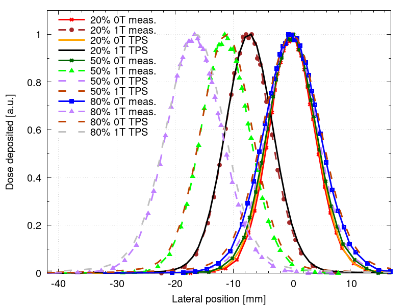
Figure 1: Normalized lateral beam profiles for a 148.2 MeV proton beam with and without an applied magnetic field of 1 T. Profiles taken at 20%, 50%, and 80% of the total range (R80).
Conclusion
The dosimetric validation of the prototype MC-based treatment planning system indicates that the effects of the magnetic field is modelled with high and sufficient accuracy. Work in progress is the evaluation of more complex heterogeneous scenarios as well as the investigation of optimization effectiveness.