Treatment plan dose calculation using the novel HyperSight CBCT
PO-1809
Abstract
Treatment plan dose calculation using the novel HyperSight CBCT
Authors: Amanda Cherpak1, Clara Fallone1, R Lee MacDonald1, Krista Chytyk-Praznik1, James L Robar1
1Nova Scotia Health & Dalhousie University, Medical Physics, Halifax, Canada
Show Affiliations
Hide Affiliations
Purpose or Objective
To compare the dose distribution of a two-arc VMAT plan calculated on images of a porcine head using HyperSight CBCT, GE Siemens kV-FBCT and Truebeam CBCT.
Material and Methods
A porcine head was imaged using the novel HyperSight CBCT imaging system installed on an Ethos linac. The head was also scanned on a GE Optima 580 RTCT fan-beam CT scanner and on a Truebeam CBCT. The standard pelvis protocol was used on all modalities to match beam quality and CTDI as closely as possible. The FBCT image was used to contour a 250 cm3 target that encompassed both bone and soft tissue. The target was located on the left lateral side of the image and extended 7 cm in the superior-inferior direction. One of the bones partially included in the target volume was also contoured as a region of interest. Contours were propagated on both HyperSight and Truebeam CBCT images through rigid image registration with the FBCT dataset. A two-arc VMAT plan was optimized and calculated on the FBCT image as per standard workflow. The plan was then forward calculated using preset monitor units on both CBCT images. The resulting dose distributions were compared to investigate the validity of using HyperSight CBCT images for dose calculation in the absence of a helical FBCT and to compare with the current standard for daily imaging, Truebeam CBCT.
Results
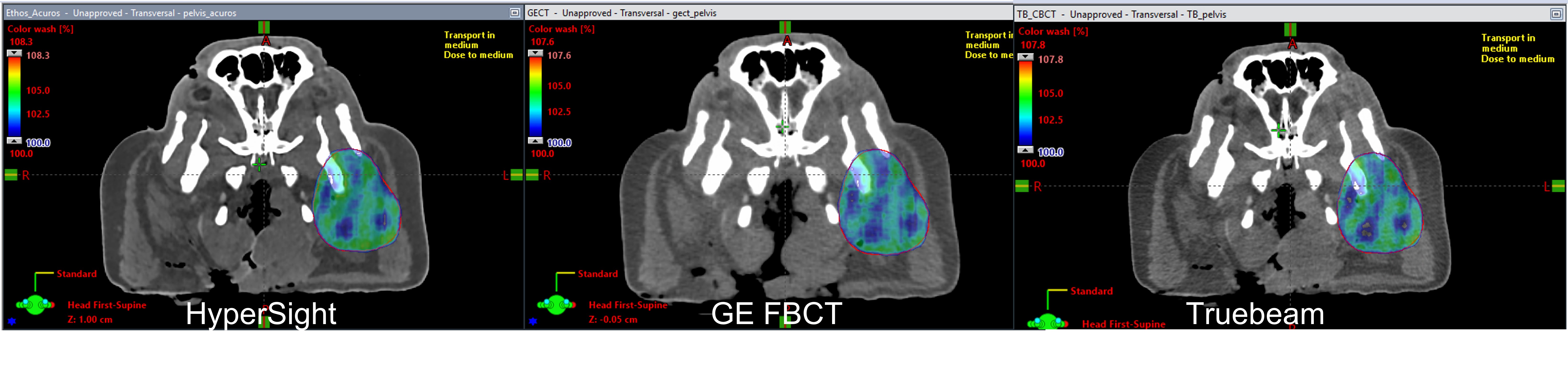
The mean dose to the target as calculated on the HyperSight image (107.6%) was within 0.2% of that as calculated on the FBCT (107.8%). The mean dose was slightly higher for the dose distribution calculated using the Truebeam CBCT (108.3%). The volume covered by 100% of the dose, V(100%), was 98.0% for FBCT, 98.9% for the HyperSight, and 96.1% for Truebeam CBCT. The loss of coverage when using the Truebeam CBCT was predominantly in the regions of bony anatomy within the target, as shown in Figure 1. The mean dose to the bone-only ROI was 92.2% for FBCT, 92.2% for the HyperSight, and 91.7% for Truebeam CBCT. Dose profiles indicate similarity amongst all three dose distributions. A 3D gamma analysis also showed small deviations between plans in the low dose region around the periphery of the body.
Conclusion
Dose calculation using the porcine head phantom resulted in similar dose distributions across all three imaging modalities. Deviations from the FBCT were found in regions of bony anatomy when using the Truebeam CBCT. This effect was not seen on the HyperSight treatment plan. This preliminary evaluation shows potential for use of HyperSight CBCT for treatment planning and dose calculation in the absence of FBCT. Inconsistencies in dose distribution were found when using the current standard for daily IGRT, Truebeam CBCT.