Validation of Beam Modeling and Dose Calculation of Proton Beam Therapy for Monaco TPS
PO-1799
Abstract
Validation of Beam Modeling and Dose Calculation of Proton Beam Therapy for Monaco TPS
Authors: Shupeng Chen1, Xiaoqiang Li1, Lewei Zhao1, Rohan Deraniyagala1, Weili Zheng1, An Qin1, Xuanfeng Ding1
1Beaumont Health, Radiation Oncology, Royal Oak, USA
Show Affiliations
Hide Affiliations
Purpose or Objective
To validate the physical beam modeling and the accuracy of proton dose calculation algorithms in Monaco treatment planning system (TPS) for proton therapy treatment.
Material and Methods
Monaco TPS proton module (ver. 6.0, Elekta, Stockholm, Sweden) Pencil Beam Scanning (PBS) and Monte Carlo (MC) dose calculation algorithms were evaluated based on the IBA ProteusONE® system. The evaluations include:1) Pristine Bragg peak proton range in water comparison between Monaco and Raystation. Mono-energetic proton beams (10x10 field) with the energy of 70MeV, 100MeV, 150MeV, 200MeV, and 227.7MeV were evaluated, respectively; 2) physical beam model verification using 9 standard cube plans that were created to simulate different treatment sites, target volumes and positions. The dose calculated in Monaco was compared with the measurements for both PBS and MC dose calculation; 3) patient-specific dose distribution comparison was performed between Monaco and Raystation. Four intensity modulate proton therapy treatment plans including 1 prostate, 1 brain, 1 lung, and 1 head and neck patient were evaluated; 4) Dose calculation accuracy was verified using the IROC phantom of standard output check and phantoms of different treatment sites including prostate, spine, lung, brain, and liver.
Results
The deviation of percentage depth dose at midSOBP was within 0.8% for PBS and 2% for MC. The point doses were measured at the depth of 5 ~ 21cm and the deviation were -1.0% ~ 1.8% for PBS and -1.2% ~ 2.4% for MC. The dose discrepancy distributions and the DVHs for the 4 patients were shown in Figure 1. The dose discrepancies between the calculated dose and the measurements of different IROC phantoms with respect to the two TPS were shown in Figure 2.
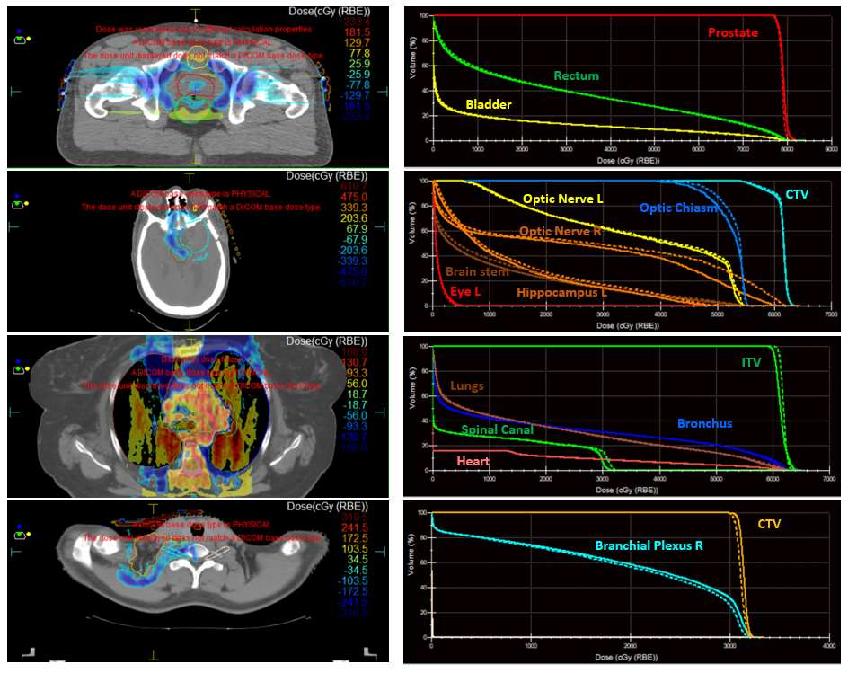
Figure 1 Spatial distributions of dose deviation between Raystation and Monaco as well as the corresponding DVHs for the 4 individual patients
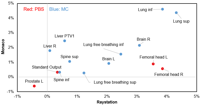
Figure 2 Deviations between the TPS calculated doses (Raystation vs Monaco) and the TLD measurements from different IROC phantoms. Abbreviations: R = right; L = left; inf = inferior; sup = superior; TLD = Thermoluminescence dosimetry
Conclusion
The physical beam modeling and proton dose calculation algorithm were precise and the associated calculation uncertainty was within clinical tolerances.