On the compaison of NIPAM gel dosimeters with high resolution MRI sequences
Melissa Cinq-Mars,
Canada
PO-1790
Abstract
On the compaison of NIPAM gel dosimeters with high resolution MRI sequences
Authors: Melissa Cinq-Mars1, Jean-David Jutras2, Luc Beaulieu3
1Université Laval, Medical Physics, Québec, Canada; 2Tom Baker Cancer Center , Physicist, Calgary, Canada; 3Université Laval, Physicist, Québec, Canada
Show Affiliations
Hide Affiliations
Purpose or Objective
The need to validate a dose distribution in three dimensions for modern three-dimensional treatment planning has generated interest in gel dosimetry. In fact, polymer gel can measure dose in 3D due to polymerization reactions occurring while the gel is exposed to radiation (De Deene et al. 2022). However, there is no agreement on an optimal recipe that has a high signal-to-noise ratio (SNR) and a large dynamic range. The aim of this project is to fine tune a NIPAM recipe based on the comparison of four recipes presented in previous studies and to characterize the spatial sensitivity and uniformity of the gel.
Material and Methods
The chosen recipes contain 1.5% (Olding et al. 2011), 3% (Senden et al. 2006), 5% (Chiang et al. 2013) and 15% (Maynard et al. 2020) of NIPAM. The recipes are compared in term of their cost, their production time and their response to dose using relaxation rate R2 (1/T2) maps. The phantoms are irradiated with 24 photon beams of 6 MV, 12 each side of the phantom in parallel-opposed configuration, with two gantry angles to produced a uniform dose distribution from 0.5 Gy to 18 Gy by changing the jaws sizes. The evaluation of the image quality with the SNR values is made and related to products cost. The total cost by SNR units at doses of 1 Gy and 10 Gy for a 1L phantom is calculated. To characterize the spatial sensitivity response, one of the phantom is irradiated with a 16 photon beams to obtain a 8 levels dose distribution from the lowest dose at the top of the phantom to the highest dose at the bottom (TB). A second phantom is irradiated, with the same beams, from the highest to the lowest dose at the top (BT). The R2 map for the SNR analysis on the 12 dose levels was obtained using the DESPOT2 mapping technique on a 1.5T Siemens Sola MRI scanner. As for the uniformity test with 8 dose levels a 16-echos CPMG sequence was employed.
Results
The results presented in figure 1 show that increasing the NIPAM concentration gives a greater dynamic range and dose can be read from 0 Gy to 13 Gy for the 15% recipe while the 1.5% recipe allows dose detection from 0 Gy to 5 Gy. The analysis of the cost in term of SNR units has shown that the recipe with 5% of NIPAM gives a better image quality-cost trade-off. Comparison of the R2 maps produced from recipes 2 and 3 (figure 2), has shown that the R2 is from 1.05 to 1.3 time higher for the phantom irradiated with the highest dose at the bottom of the jar (TB). We believe to be a gravity-dependent settling for the polymer chains, as hypothesized previously in a CT-based NIPAM gel analysis by Maynard et al. 2020.
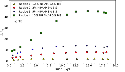
Figure 1 – ∆R2 values as a function of dose for the irradiation from top to bottom (TB).
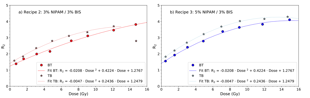 Figure 2. R2 according to dose for recipes 2 and 3.
Conclusion
The next step of the project is to manufacture phantoms of an optimized recipe. The dynamic range, the response to dose and the sensibility of the gel will be characterize, as well as a method for correcting the non-uniform sensitivity response in three dimensions.