Modelling digital anthropomorphic phantoms for upright position treatment studies
Wioletta Kozłowska,
Switzerland
PO-1747
Abstract
Modelling digital anthropomorphic phantoms for upright position treatment studies
Authors: Wioletta Kozłowska1,2, William Larsen1,2,3, Janine Bossé2, Paul Segars4, Jonathan Farr1,2
1Applications of Detectors and Accelerators to Medicine SA, Clinical Office, Meyrin, Switzerland; 2Advanced Oncotherapy plc, Clinical Office, London, United Kingdom; 3Ecole Polytechnique Federale de Lausanne (EPFL), Microengineering, Lausanne, Switzerland; 4Duke University , Radiology, Durham, USA
Show Affiliations
Hide Affiliations
Purpose or Objective
Upright radiotherapy treatment using a chair for patient positioning brings advantages in cost and facility space reduction, as well as allowing the practical implementation of particle arc therapy. In addition, certain treatment indications may benefit from smaller diaphragm movements [1] and changes in internal organ positioning, due to gravitational force. However, as vertical imaging systems are limited, it is challenging to properly evaluate changes to human anatomy that could affect upright radiotherapy indications. To facilitate treatment planning studies using an upright positioning system, we propose the use of digital anthropomorphic XCAT2 phantoms [2], modified to the seated position, with simulated and validated changes of the internal anatomy.
Material and Methods
Four XCAT2 phantoms were modified from the supine to upright position, with a backrest angle of 20˚, using internal XCAT scripts and Rhino 7 (Robert McNeel & Associates, USA). Subsequently, organ translations and deformations in the abdominal and pelvic areas, resulting from gravitational force, were simulated for selected organs using Finite Element Method (FEM) simulations with ANSYS 19 software (ANSYS Inc., USA). Finally, models were reassembled, with each organ in the phantom assigned a respective HU value, and virtual DICOM CT files were created for treatment planning studies.
Results
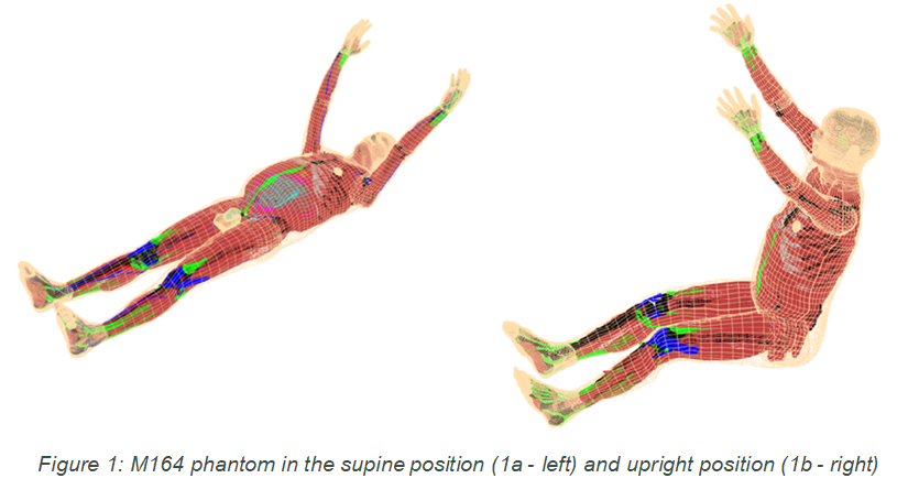
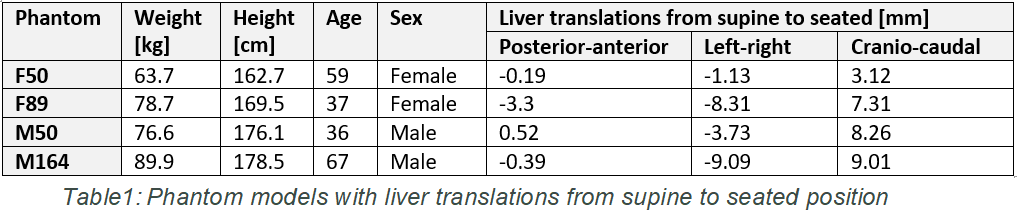
Figure 1 presents a male XCAT2 phantom in supine (1a) and upright (1b) positions. The FEM studies showed a small displacement of the abdominal and pelvic organs, comparable with results found in literature [3]. Table 1 summarizes data from patient models and provides example information on translation of the liver organ from the supine to the seated position using FEM studies. Our results showed average liver translations of -0.84 mm, -5.6 mm, and 6.92 mm in the posterior-anterior, left-right, and cranio-caudal directions, respectively. For the left-right and anterior-posterior directions, our averaged results differ by 44% and 20% respectively from those reported by others [3]. However, the cranio-caudal direction showed a 3x smaller translation compared to the single subject results presented in [3]. This discrepancy may be due to the simplified model used for liver ligaments during our FEM studies, and this is currently under investigation.
Conclusion
Our results indicate that anthropomorphic digital phantoms can be valuable tools in treatment planning optimisation for upright positioning geometries. Future benchmarking and refining work will be performed using additional data from volunteers’ CT and ultrasonography scans for statistical analysis. In addition, we plan to model lung and diaphragm motions in the upright positioned phantoms during breathing process.
[1] Takazakura et al. 2014. J Magn Reson Imaging. 19:605-9.
[2] Segars et al. 2010 MedPhys 37(9) :4902-4915
[3] Hayes et al. 2013. Comput Math Methods Med. 2013:419821