Multicentric characterisation of lateral beam profiles from 6 MV FFF beam of three 0.35T MR-linacs
Antonella Fogliata,
Italy
PO-1744
Abstract
Multicentric characterisation of lateral beam profiles from 6 MV FFF beam of three 0.35T MR-linacs
Authors: Antonella Fogliata1, Randa El Gawhary2, Davide Cusumano3, Lorenzo Placidi4, Flaviovincenzo Quaranta3, Matteo Nardini4, Maria Rago2, Luca Indovina4, Sebastiano Menna3
1Humanitas Research Hospital IRCCS, Radiotherapy, Rozzano-Milan, Italy; 2San Pietro FBF Hospital, Radiotherapy, Rome, Italy; 3Mater Olbia Hospital, Radiotherapy, Olbia, Italy; 4Fondazione Policlinico Universitario Agostino Gemelli IRCCS , Radiotherapy, Rome, Italy
Show Affiliations
Hide Affiliations
Purpose or Objective
The advent of MR-guided radiotherapy technologies, combining flattening filter free (FFF) linear accelerators with on-board MR scanners, opened new clinical opportunities and physical challenges. The physical characterisation of FFF beam profiles in presence of a magnetic field is a new and relevant topic in the field of radiation dosimetry, for which new standardisation procedures and formulation are needed.
The aim of this multicentric study is to propose new normalisation factors by analysing such lateral beam profiles, to allow for the calculation of standard parameters typical of flattened beams, such as penumbra and symmetry.
Material and Methods
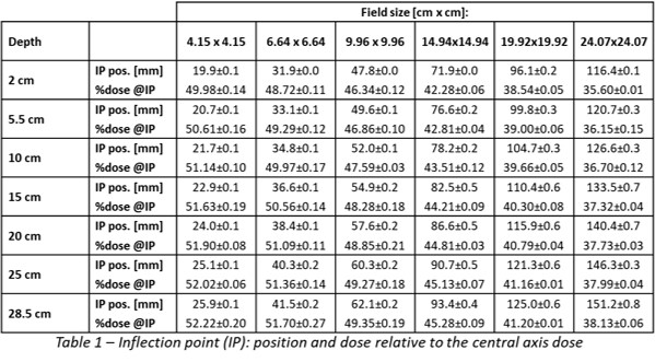
The experimental measurements were carried out on three 0.35 T MRgRT MRIdian (ViewRay) systems. In all the Institutions an equal set of measurements was acquired using the same equipment, consisting of a 3D motorised water phantom (Tales 3D MR, LAP) and a 0.125 cc vented ion chamber with an inner diameter of 5.8 mm (Exradin A28MR). Lateral profiles, in the direction perpendicular to the magnetic field, of the 6 MV FFF beam were measured with fixed fine step resolution at seven different depths: 2, 5.5, 10, 15, 20, 25 and 28.5 cm. For each depth, six beam dimensions were acquired: 4.15x4.15, 6.64x6.64, 9.96x9.96, 14.94x14.94, 19.92x19.92 and 24.07x24.07 cm2. All the measurements were carried out at a source-surface distance (SSD) of 85 cm. The integration time was adjusted to balance the time for measuring and the noise reduction, and a reference detector was used during all the experimental measurements.
A post-processing procedure consisted of data smoothing within the in-field region, beam centering and mirroring. The position of the inflection point, minimum and maximum of the second derivative in the fall-off region, and the extremes of the third derivative were calculated. The profile normalization was determined by imposing the 50% dose level at the inflection point. The fitting parameters describing the renormalization considering the following formula were estimated:
Renorm=(a+b∙FS+c∙depth)/(1+d∙FS+e∙depth)
where FS is the field (for square fields) in cm, depth is the measuring depth in mm, and a, b, c, d, e are the fitting parameters.
Results
The position of the inflection point averaged over the three datasets, for all the fields and depths, is reported in Table 1, together with the percentage dose value relative to the central axis at the same inflection point.
The values of the Renormalization fitting parameters are here reported: a=94.13±0.55, b=-0.308±0.155 cm-1, c=-0.033±0.096 cm-1, d=-0.016±0.001 cm-1, e=0.002±0.001 cm-1.
Conclusion
In this multicentric study, we effectively determined and reported the fitting parameters for profile normalization of a 6 MV FFF beam from an MRIdian MR-linac in the presence of a 0.35T magnetic field. With these parameters, it is possible to calculate penumbra, unflatness and symmetry for all square field and depth combinations.