Comparing intra- and inter-fraction motion of tumors with fiducial markers for lung SBRT
PO-1339
Abstract
Comparing intra- and inter-fraction motion of tumors with fiducial markers for lung SBRT
Authors: Yuki Manabe1, Takehiro Shiinoki1, Koya Fujimoto1, Kazushi Ueda1, Masako Karita1, Miki Kajima1, Hidekazu Tanaka1
1Yamaguchi University, Radiation Oncology, Ube, Japan
Show Affiliations
Hide Affiliations
Purpose or Objective
At our institution, respiratory-gated SBRT (RG-SBRT) for lung tumors is performed using a real-time tumor monitoring system. Several fiducial markers (FMs) are inserted near the tumor for RG-SBRT. However, optimal FM as an internal surrogate cannot be selected to perform RG-SBRT. This study aimed to evaluate the relationship between FM and lung tumors to determine the optimal FM.
Material and Methods
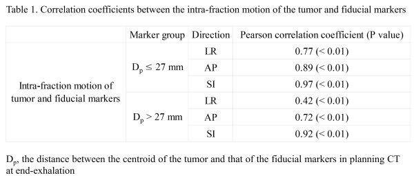
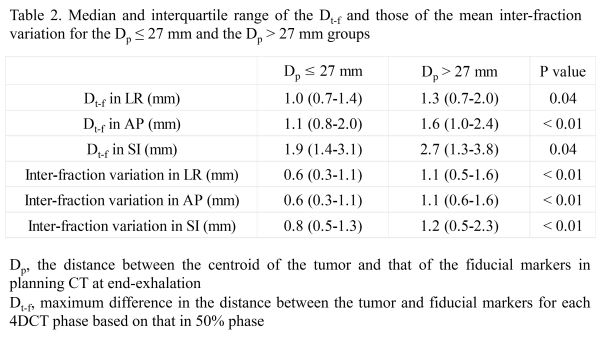
This study enrolled 41 patients who underwent RG-SBRT with real-time tumor monitoring for lung cancer. For each patient with 1-5 FMs, CT at end-exhalation (CTp) and 4DCT were acquired for treatment planning. CT at EE before treatment (CTfr) was also acquired. The dose prescription was 48-60 Gy/4-8 fr, and 145 FMs were analyzed. The FMs were divided into two groups based on the median distance between the centroid of the tumor and that of the FMs in CTp (Dp). First, to evaluate the relationship between the intra-fraction motion of the tumor and FMs during respiration, the intra-fraction motion was defined as the translation of the tumor and FMs between each phase and the 50% phase. The intra-fraction motion of the tumor and FMs were calculated in the left-right (LR), anterior-posterior (AP), and superior-inferior (SI) directions, respectively. Pearson correlation coefficients between the relative coordinates of the tumor and FMs were calculated in each direction. Second, to evaluate the positional relationship between the tumor and FMs during respiration, the distance between the tumor and FMs was calculated in each direction for each 4DCT phase, and the maximum difference in distance was calculated based on the distance between tumor and FMs in the 50% phase (Dt-f). Third, to evaluate the inter-fraction variation of the tumor using the FM setup, the CTfrs were registered to CTp using each FM for patient setup. The differences between the tumor centroids in CTp and CTfr were defined as the inter-fraction variation and the mean inter-fraction variation was calculated in each direction.
Results
The median Dp was 27 mm. For the Dp ≤ 27 mm group, the intra-fraction motion of the FMs was significantly and strongly correlated with that of the lung tumor in all directions. However, for the Dp > 27 mm group, the intra-fraction motion of the FMs was significantly and strongly correlated with that of the lung tumor only in the AP and SI directions. In the LR direction, the intra-fraction motion of the FMs was significantly and moderately correlated with that of the lung tumor (Table 1). The Dt-f for the Dp ≤ 27 mm group was significantly smaller than that for the Dp > 27 mm group in all directions. The inter-fraction variation for the Dp ≤ 27 mm group were significantly smaller than that for the Dp > 27 mm group in all directions (Table 2).
Conclusion
The fiducial marker for Dp ≤ 27 mm may improve the accuracy of RG-SBRT in terms of intra- and inter-fraction variation. Further research is needed to determine the accuracy of FMs as internal surrogates.