Determining treatment margins in hypo-fractionated (Fast Forward) breast treatment.
Frank Van den Heuvel,
Switzerland
PO-1880
Abstract
Determining treatment margins in hypo-fractionated (Fast Forward) breast treatment.
Authors: Linda van der Heijden1, Patricia Brouwers2, Bleddyn Jones3, Barbara Wachters2, Frank Van den Heuvel4,3
1Zuidwest Radiotherapeutisch Instituut,, DPK, Vlissingen, The Netherlands; 2Zuidwest Radiotherapeutisch Instituut,, Clinic, Vlissingen, The Netherlands; 3University of Oxford, Oncology, Oxford, United Kingdom; 4Zuidwest Radiotherapeutisch Instituut, Physics, Vlissingen, The Netherlands
Show Affiliations
Hide Affiliations
Purpose or Objective
A reduced number of
fractions for breast cancer treatment is indicated . Using five fractions has been shown to
be safe. The techniques used, were direct translations
of the standard, the only change was the
number and size of the various fractions.
However, margins used with a high number of fractions were used, but the statistical power of the various clinical trials
was likely to be insufficient to detect any inadequacies.
1. Margin
calculations rely on the concepts of normal distributions, an invalid assumption.
2. Margins impact healthy tissue, which can vary depending on the
radio-biological properties of specific normal tissues
Here we analyse the efficacy of the current margins
used in whole breast treatments.
Material and Methods
20 patients (10 left, 10
right) treated to the whole breast were entered in an audit study. Plans
were generated for treatments 2.67Gy x 15 frx and 5.2Gy x 5 fx, using the recommended
constraints for normal tissue.
For
each patient 20 systematic errors are generated, and
doses recalculated. Shifts shifts from this position is determined by the number of fractions.
For each treatment instance the Effective Uniform Dose (EUD) for all structures is calculated. Here we report on Heart, Lung ( α/β = 3.0
late toxicity) and Liver ( α/β = 1.0).
Results
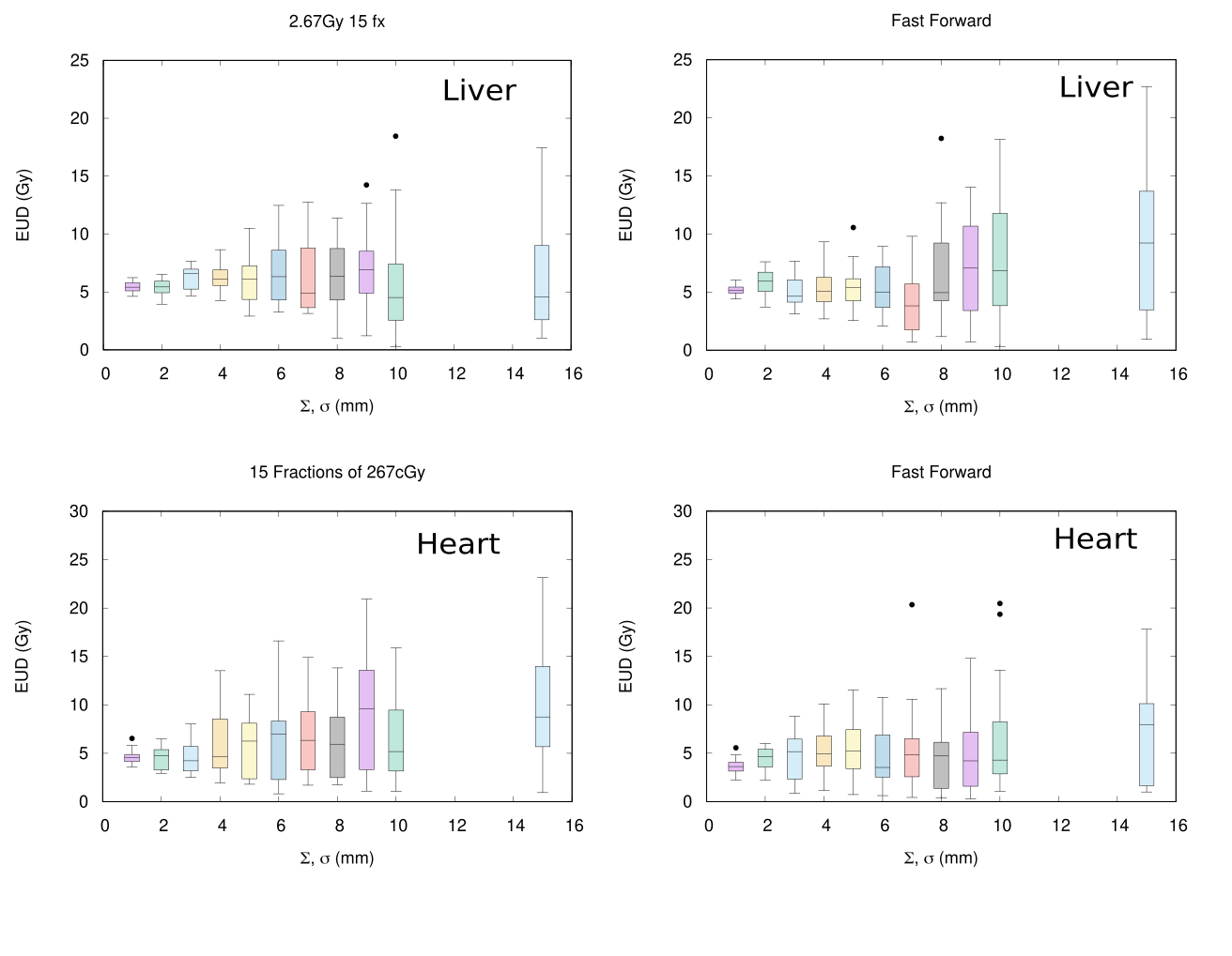
Figure 1 shows the variations of EUD (Gy)
in a boxplot as a function of the systematic and random errors
(Σ and σ) .
In
the fast forward arm more outliers are noted indicating
“unlucky” patients. However the outliers only occur with Σ and σ larger than 5mm.
Conclusion
Margins used in the treatment of breast
cancer are adequate if errors are
smaller than 5mm. Daily treatment imaging becomes a necessity in order to achieve this
degree of accuracy.