Reproducibility of diffusion-weighted magnetic resonance imaging in head and neck cancer
PO-1750
Abstract
Reproducibility of diffusion-weighted magnetic resonance imaging in head and neck cancer
Authors: Jonas Habrich1, Victor Fritz2, Simon Böke3, Marcel Nachbar1, Konstantin Nikolaou4, Fritz Schick2, Daniel Zips3, Daniela Thorwarth1
1University Hospital and Medical Faculty, University of Tübingen, Section for Biomedical Physics, Department of Radiation Oncology, Tübingen, Germany; 2University of Tübingen, Section for Experimental Radiology, Department of Diagnostic and Interventional Radiology, Tübingen, Germany; 3University Hospital and Medical Faculty Tübingen, Department of Radiation Oncology, Tübingen, Germany; 4University Hospital Tübingen, Department of Diagnostic and Interventional Radiology, Tübingen, Germany
Show Affiliations
Hide Affiliations
Purpose or Objective
MR-Linacs (MRL) allow for sequential acquisition of functional magnetic resonance imaging (MRI) and therefrom derived quantitative imaging biomarkers such as apparent diffusion coefficients (ADC). The prognostic value of ADC derived from diffusion-weighted (DW)-MRI has been shown in offline studies in head and neck cancer (HNC), but for application at MRL, technical validation of DW sequences is required first.
The aim of this study was to assess the reproducibility of ADC values derived from DW-MRI in HNC patients acquired with a hybrid 1.5 T MRL compared to a 3 T diagnostic scanner (DS).
Material and Methods
Eight patients with locally advanced HNC were treated at the 1.5 T MRL (Unity, Elekta) and examined with DW-MRI at the MRL and additionally at a 3 T diagnostic scanner (Vida, Siemens) before (measurement 1) and during week 2 (measurement 2) of radiotherapy. ADC maps were calculated using a pixel-wise mono-exponential fit to signal amplitudes of the b150- and b500 images of the MRL and b200-, b500 and b1000 images of the 3 T scanner. Details of the sequence parameters are presented in Table 1. Mean ADC values and their relative differences were calculated for gross tumor volume (GTV), submandibular and parotid glands. Bland-Altman analysis with limits of agreement (LoA, ± 1.96·standard deviation) was performed to investigate reproducibility.
Table 1: Details of imaging protocols
| MRL
| DS
|
TE/TR [ms]
| 68/4811
| 44/10800
|
Acquisition voxel size [mm³]
| 3x3x4
| 3x3.3x5
|
Acquisition matrix
| 132x134x25
| 128x76x24
|
Flip angle
| 90°
| 90°
|
Bandwidth [Hz/px]
| 1930
| 2298
|
b-values (averages) [s/mm²]
| 150 (5), 500 (8)
| 200 (6), 500 (7), 1000 (10)
|
Results
A total of 13 measurements were available for analysis. Mean average ADC values for GTV, submandibular and parotid glands for the MRL were 1251, 1114 and 789·10-6 mm2/s and for the DS those were 1483, 1347 and 1038·10-6 mm2/s. Figure 1 shows Bland-Altman plots for GTV, submandibular and parotid glands. Mean absolute ADC differences ± LoA for GTV, submandibular and parotid glands were 232±430, 234±351 and 249±302·10-6 mm2/s. Accordingly, mean relative differences resulted in 15%, 17% and 24%, respectively.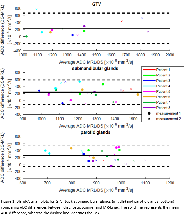
Conclusion
This study showed good overall reproducibility of ADC measured at the MRL with respect to a DS. However, comparison of ADC values showed an underestimation of the ADC for the MRL. This shift must be considered when results from DS are translated to the MRL and vice versa. Further validation with more patients is necessary to confirm these results.