Accuracy of DIR-based intra-fraction motion management in MR-guided radiotherapy
Miguel A. Palacios,
The Netherlands
PO-1695
Abstract
Accuracy of DIR-based intra-fraction motion management in MR-guided radiotherapy
Authors: Miguel A. Palacios1, Georgi Gerganov2, Paul Cobussen1, Shyama U. Tetar1, Tobias Finazzi1, Berend J. Slotman1, Suresh Senan1, Cornelis J.A. Haasbeek1, Iwan Kawrakow2
1Amsterdam UMC - VUmc location, de Boelelaan 1117, 1081 HV, Department of Radiation Oncology, Amsterdam, The Netherlands; 2Viewray Inc., 2 Thermo Fischer Way, Science Department, Oakwood Village, Ohio 44146, USA
Show Affiliations
Hide Affiliations
Purpose or Objective
MR-guided radiotherapy (MRgRT) enables real-time monitoring of the
anatomy of the patient during treatment delivery. Manual delineations for tumor
contour tracking during delivery is impractical and very labor intensive. In
this work we studied the accuracy of automatic tumor segmentation based on deformable image registration (DIR) in the delivery of
MRgRT by comparing it to manual delineations performed by experienced
observers.
Material and Methods
Twenty
patients with lung, pancreatic, renal and adrenal tumors previously treated
with SBRT using MRgRT were included in the study. Patient and treatment fractions
included for this analysis were randomly selected. Five observers with at least
2 years of experience in MRgRT delineated the gross tumor volume (GTV) for 20
patients on 240 frames of an MR-cine on a sagittal plane, with 0.35cm x 0.35cm
in-plane resolution. DIR-based GTV
contours were propagated using 4 different algorithms from a reference frame to
subsequent frames, in order to assess the accuracy of online DIR-based contour
tracking. Each DIR-algorithm was implemented as a combination of several
independent tracking modules, referred to as “trackers”.
Geometrical
analysis based on the Dice Similarity Coefficient (DSC), centroid distance and
Hausdorff Distance (HDD) were performed to assess the inter-observer
variability and the accuracy of automatic segmentation. A Confidence Value
metric for the reliability of the tumor auto-contouring was also calculated.
The Confidence Value is based on the correlation of the image registration and
intersection and union of the results from each of the trackers composing the
algorithms.
Results
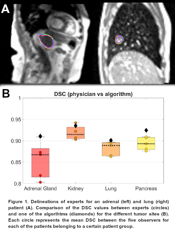
Figure 1A shows the delineations of the
experts for an adrenal and lung case. Inter-observer delineation variability
resulted in mean DSC, HDD and centroid distance among observers across all
patients of 0.89, 5.8 mm and 1.7 mm, respectively. Figure 1B shows the
comparison for the DSC values between the experts and one of the algorithms for
all tumor sites. The four DIR algorithms were able to reproduce the original
contour delineated by each of the observers on the reference frame to
subsequent frames with an excellent agreement. Mean DSC for each algorithm
across all patients and frames was > 0.90, whereas the HDD and centroid
distances were below 4.0 mm and 1.5 mm, respectively. All four algorithms
performed equally good independently of the specific tumor site. The Confidence
Value was linearly correlated with the DSC.
Conclusion
DIR-based auto-contouring in MRgRT
exhibited a high level of agreement with the manual contouring performed by
experts. In addition, DIR-based auto-contouring resulted in a lower variability
across all quantitative metrics compared to the inter-observer variability. Clinical
implementation of DIR-based algorithms for intra-fraction motion correction in
MRgRT provides clinicians and physicists with robust and reliable tools to
deliver radiation dose accurately.