MR-guided adaptive radiotherapy for gastric lymphomas: a pilot study to select PTV margins
PO-1690
Abstract
MR-guided adaptive radiotherapy for gastric lymphomas: a pilot study to select PTV margins
Authors: Laura Rechner1, Kristian Boye1, Peter Meidahl Petersen1
1Copenhagen University Hospital - Rigshospitalet, Department of Oncology, Center for Cancer and Organ Diseases, Copenhagen, Denmark
Show Affiliations
Hide Affiliations
Purpose or Objective
Gastric lymphoma is a tumor site that is
very difficult to visualize on CBCT. This results in difficulty in using CBCT
for both image matching for setup and the evaluation of daily anatomical variations
of the stomach, which can be significant. Therefore, adaptive radiotherapy on
the MR-linac could benefit patients with gastric lymphoma by providing high
soft tissue contrast for setup, gating on internal anatomy, and the possibility
to adapt to the daily anatomy. However, appropriate PTV margins to account for
intra-fraction variations for this tumor site are unknown. Therefore, the
purpose of this work was to evaluate the intra-fraction variation in the
position of the stomach in a few pilot cases and to establish a workflow for
determining patient-specific PTV margins for adaptive radiotherapy of the
stomach on the MR-linac.
Material and Methods
Two gastric lymphoma patients and one
healthy volunteer underwent simulation on the MR-linac (MRIdian, ViewRay). Simulation
MRIs were acquired in inspiration breath hold at two separated timepoints within
the same session (MRI1 and MRI2) to represent the changes that could happen
intra-fractionally, and sagittal cine images and tracking were performed after
each MRI to test the feasibility of tracking. The CTV was contoured offline on
both scans (CTV1 and CTV2) using a commercial TPS (Eclipse, Varian Medical
Systems). Registration of the MRIs was performed with focus on the region of
the target used for tracking on the sagittal cine images. PTVs were created
using expansions ranging from 5-10 mm from CTV1. CTV2 was copied to the MRI1
structure set, and it was assessed if the PTVs created from CTV1 were
sufficient to also cover CTV2.
Results
Gating on the cine images was successful
with tracking on the superior part of the stomach (Figure 1) to simulate gating
during treatment. The average time between scans was 21 minutes (range 15-33). CTV
volumes ranged from 397 to 592 cc. All three cases had complete coverage of
CTV2 by the a PTV expansion from CTV1 of 8 mm (Table 1). Furthermore, the
superior region of CTV2 near the diaphragm was always covered by a 5 mm
expansion (Figure 2), which was expected due to focus on that region during
registration.
Figure 1
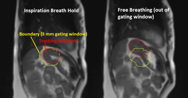
Figure 2
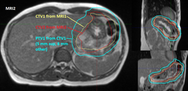
Table 1
| Volume (cc) of CTV2 outside PTV1 (expanded from CTV1)
|
|
|
| PTV margin (mm) | Patient 1 | Patient 2 | Patient 3 |
| 5 | 0.78
| 0.27
| 0.05
|
| 6 | 0.47
| 0.04
| 0.00
|
| 7 | 0.02
| 0.00
| 0.00
|
| 8 | 0.00
| 0.00
| 0.00
|
| 10 | 0.00
| 0.00
| 0.00
|