The effect of high density material in breast expanders on the dose distribution
PO-1590
Abstract
The effect of high density material in breast expanders on the dose distribution
Authors: Leen Paelinck1, Chris Monten1, Carlos De Wagter1, Yolande Lievens1
1University Hospital Ghent, Radiotherapy, Ghent, Belgium
Show Affiliations
Hide Affiliations
Purpose or Objective
A breast expander is a temporal prosthesis
implanted in the breast during mastectomy and consists of a magnet, injection
port and an expansion envelope. The high densities of the magnet and injection
port have an influence on the image quality and the accuracy of dose
calculations. The purpose of this study was to investigate which density
overrides are appropriate to use in our TPS Raystation 6 (RaySearch, Stockholm,
Sweden) by comparing calculations and radiochromic film (Gafchromic, Ashland
Specialty Ingredients, USA) measurements.
Material and Methods
A schematic representation of the
measurement setup through a (non-isocentric) transversal plane is shown in
figure 1. The breast expander with a filled expansion envelope was immersed in
a polystyrene box filled with water and placed on the top of a slabbed
polystyrene phantom. Additional polystyrene plates were placed next to the
water filled box. This allows the placement of radiochromic films in a coronal
and sagittal plane just under and next to the box. Two beam setups, G = 0° and
G = 90°, were used. The beam size and film size fitted the dimensions of the
box in the coronal and sagittal measurement planes. Each time 200MU was
delivered. A calibration curve was measured and the film analysis was performed
by in-house made software. A CT scan (Aquilion, Toshiba Medical
Systems, Tokyo, Japan) of the phantom setup was made and imported in the TPS.
The contouring of the magnet and port were respectively based on a L/W of
6000/10000 and 2000/1000. Four density
override configurations were investigated: 1) magnet and port on titanium, rest
of the phantom on water, 2) magnet on titanium, port and rest of the phantom on
water, 3) no override on magnet and port, rest of the phantom on water and 4)
magnet on titanium, port on aluminum, rest of the phantom on water. In all cases,
the predefined materials in RayStation ‘water’, ‘titanium’ and ‘aluminum 2’ were
used. All calculations were performed with a collapsed cone algorithm.
Results
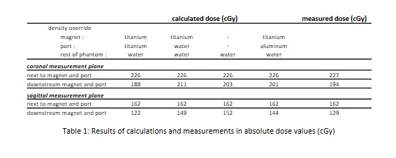
In figure 1 an image of the measured dose in
both orientations is shown. The attenuation caused by the magnet and port is
clearly visible on both films. In table 1 the results of the calculations and
measurements in the homogenous region a few cm away from the magnet and port
and just downstream the magnet and port are displayed in absolute dose values
(cGy). The maximum measured attenuation caused by the magnet in the sagittal
and coronal measurement plane is respectively 20% and 15%. The case in which
magnet and port are overridden with density titanium and the rest of the
phantom with water is in best agreement with the calculations for both measurement
orientations.
Conclusion
This study investigated how to deal with
the presence of breast expanders in the radiotherapy treatment process. The
high density materials affects the image quality and accuracy of dose
calculations. This study shows that a density override of magnet and port with
titanium is in best agreement with radiochromic film measurements.