Dosimetry standardization of Total Body Irradiation of mice in a SARRP irradiator
Ileana Silvestre Patallo,
United Kingdom
PO-1584
Abstract
Dosimetry standardization of Total Body Irradiation of mice in a SARRP irradiator
Authors: Ileana Silvestre Patallo1,2, Feiyan Shi2, Rebecca Carter3, Andy Nisbet2
1National Physical Laboratory, Medical Radiation Physics, Teddington, United Kingdom; 2University College London (UCL), Medical Physics and Biomedical Engineering, London, United Kingdom; 3University College London (UCL), Cancer Institute, London, United Kingdom
Show Affiliations
Hide Affiliations
Purpose or Objective
Total Body Irradiation (TBI) of mice is used to generate models of
diseases such as leukaemia, by suppressing the haematopoietic system prior to systemic
tumour cell engraftment. Such models can be used to evaluate the outcomes
of different therapeutic methods.
Dosimetry of TBI in image guided preclinical irradiators is challenging.
The main problem arises from the variety of pre-clinical irradiation conditions
and the difficulties in relating those to the recommendations from well-established
clinical dosimetry protocols in the range of medium energy x-rays.
In
this study, a methodology for accurate and standardized deliveries of dose to
mice during TBI was experimentally determined. Dose across different regions of
the mouse and the effect of the mouse positioning system were assessed. As a
consequence, a reproducible irradiation protocol, for the use of the Small
Animal Radiation Research Platform (SARRP), was established.
Material and Methods
The SARRP’s reference dosimetry was performed with the local secondary
standard system with a calibration traceable to the National Physical Laboratory’s
Air Kerma primary standard. Measurements were performed in a solid water slab
phantom following manufacturer’s recommendations. Alanine verification of the
output was determined, in the same conditions of irradiation of the ionization
chamber measurements. [1].
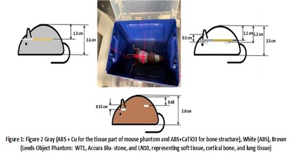
EBT3 Gafchromic film and alanine pellets were subsequently used to
measure dose delivered to three mice phantoms, mimicking positioning of real
mice during TBI irradiations. The mice phantoms were different in their material
composition [2] and the type of detectors that they were able to hold: film,
alanine or both (Figure 1). Irradiation times to film and alanine were
different, considering that alanine uncertainties are lower for dose over 10
Gy.
Results
Dose to film in the abdominal region of the Gray and
White mice phantoms compared within 3.2%. Uncertainty based on the standard
deviation of the mean (SDOM) of three independent repetitions was 2.3% Dose to
alanine in the abdominal region of the Brown and White phantoms agreed within
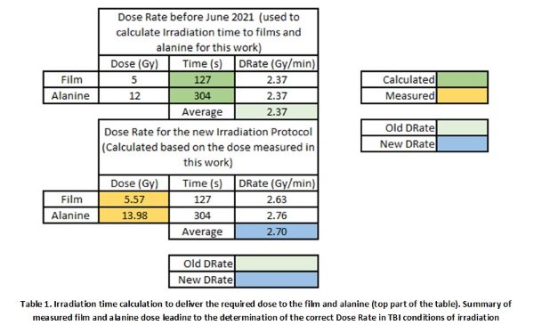
2.1% with 0.6% uncertainty. Film and alanine dose at approximately the same
position in the abdominal region of the White phantom, agreed within 2%. On
average (both detectors and three phantoms), the dose rate (DR) (measured dose/irradiation
time) to a point in the mid plane of the mouse in the abdominal region was 2.7
Gy/min. The DR currently used to calculate TBI irradiation times within our
institute (based on 2018 measurements with an inadequate dosimetric system) was
2.37 Gy/min.
Conclusion
Previous to this work, the dose delivered during TBI
irradiations was 13% larger than required. The DR previously used to calculate
irradiation times did not consider the differences between reference output
measurement conditions and real setup of mice during TBI experiments. A standardized
protocol with a detailed description of the irradiation conditions and table
for time calculation is now routinely used.