Characterization and the image quality of 2.5 Megavoltage Beam in TrueBeam linear accelerator.
PO-1541
Abstract
Characterization and the image quality of 2.5 Megavoltage Beam in TrueBeam linear accelerator.
Authors: Hubert Szweda1, Bartosz Pawałowski1, Olga Bąk2, Katarzyna Paciorkowska1, Krzysztof Matuszewski2, Maja Rumak3
1Greater Poland Cancer Centre, Department of Medical Physics, Poznań, Poland; 2Greater Poland Cancer Centre, Department of Medical Physics , Poznań, Poland; 3University of Medical Sciences, Electroradiology Deparment, Poznań, Poland
Show Affiliations
Hide Affiliations
Purpose or Objective
Over the past three decades, electronic imaging
portal devices became a very important tool used for pretreatment patient
verification and one of the most useful devices in the patient specific QA in
radiotherapy. Potentially, using kilovoltage imaging offers better contrast of
soft tissues, but the quality of the megavoltage imaging has been constantly improved.
In the newest version of Varian TrueBeam medical accelerators, the energy of MV
imaging beam has been reduced from 6MV to 2.5MV. New beam generated by 2.5MV
nominal accelerating potential reduces the dose of radiation received by the
patient during the imaging process by 50% and provides significantly better
image quality. During the study we performed characterization of the 2.5MV beam
by measuring percent depth-dose, beam profiles and outputs. To control the
quality of the imaging we used TOR 18FG phantom for low‐contrast detectability and spatial resolution.
Material and Methods
In order to measure PDD, Roos
(type 34001) ionization chamber was used. To perform measurement of beam
profiles, we used Semiflex 3D (type 31021) and to calculate outputs we used
Farmer chamber (type 30013). Second method of dose verification was accomplished with EBT3 radiochromic films.
We performed Winston-Lutz Test to check accordance
of 2.5MV and 6MV isocentres. To evaluate image quality, TOR 18FG
phantom was used. To simulate scatter effect in the patient body, slabs (5cm and 10 cm) of solid water were
positioned on the beam path.
Results
We characterized the 2.5MV beam by evaluating Dmax
(6.01mm), D100mm (51.52%), D200mm (21.83%), dose on
surface (95.89%), and quality index (0.4770). In regard to control beam profiles
for 40x40cm2 field, we checked profiles intensity for distances ± 60mm and ± 180mm from the central beam axis. Obtained
values were 96.2% and 73.3% respectively.
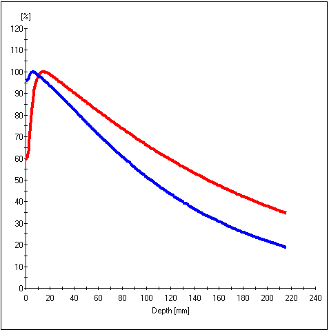
Fig.1 - Comparison between percent depth-dose curves of 2.5MV (blue curve) and 6MV (red curve).
We observed good agreement between radiation
isocenter for 2.5MV and 6MV. In regard to imaging quality, 2.5MV showed much
better image quality compared to the traditional MV beam. With 2.5MV
beam it was possible to detect all low-contrast objects without any solid water
layer and detect 13 more objects than 6MV beam for 10cm solid water layer. Megavoltage
beam with reduced energy also demonstrated better spatial resolution.
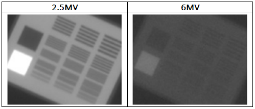
Fig.2 - Comparison between image quality of 2.5MV and 6MV for TOR 18FG phantom, without any layer of solid water on the phantom.
Conclusion
This work characterized the 2.5MV beam used for
patient imaging in the TrueBeam medical linear accelerators. The 2.5MV beam showed a much better imaging
quality compared to the conventional 6MV energy. A significant advantage of the 2.5MV beam is
the reduction of the imaging dose received by the patient during a radiotherapy
session. Evaluated parameters describing the quality of
the beam were used to develop quality control protocols for clinical practice.