Radioluminesence imaging for CyberKnife® dosimetry and quality assurance
Antonello Spinelli,
Italy
PO-1525
Abstract
Radioluminesence imaging for CyberKnife® dosimetry and quality assurance
Authors: Antonello Spinelli1, Zahra Shakarami1, Sara Broggi2, Antonella del Vecchio2, Claudio Fiorino2
1San Raffaele Scientific Institute, Experimental Imaging Center, Milan, Italy; 2San Raffaele Scientific Institute, Medical Physics, Milan, Italy
Show Affiliations
Hide Affiliations
Purpose or Objective
To investigate the applicability of
radioluminescence imaging (RLI) as a novel 2D quality assurance (QA) dosimetry
system for CyberKnife®.
Material and Methods
We developed a real time dose measurement system
based on a commercial complementary metal oxide semiconductor (CMOS) camera
facing a radioluminescence screen located at the isocenter (Figure.1). The
radioluminescence light collected by a lens was used to measure 2D dose
distributions. An image transformation procedure, based on two reference
phantoms (square and star), was developed to correct for projective distortion
due to the angle (15 degrees) between the optical and the radiation beam main axis.
Dose profiles were measured for field sizes ranging from 10 mm to 60 mm using
fixed circular and Iris™ collimators and compared against gafchromic film
(GC). The corresponding full width at half maximum (FWHM) was measured using
the RLI and benchmarked against GC film. Small shift of the isocenter position were introduced on purpose to test
the sensitivity of the RLI system to field size variations.
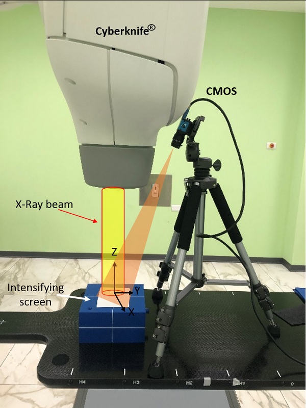
Figure 1: RLI acquisition
setup. The radiation beam (yellow) impinged on
the
scintillation screen, which then emits light collected by the camera (orange).
Results
The FWHM measurements using the RLI system
indicated strong agreement with GC film with maximum absolute difference equal
to 0.131 mm for fixed collimators and 0.049 mm for the Iris (Table.1). A 2D
analysis of RLI with respect to GC film indicated that the differences in the
central region are negligible, while small discrepancies are in the penumbra
region (30%-70%). Changes in field sizes of up to 0.2 mm were detectable by RLI.
Table 1: FWHM (mm) obtained from the RLI line profiles and
compared with the GC films.
| Field Size (mm) | FWHM (mm) |
| Discrepancy |
|
| RLI | GC Film | mm | % |
| X-direction |
|
|
|
|
| Circle 10 | 10.039 | 10.108 | -0.069 | -0.68 |
| Circle 40 | 41.206 | 41.269 | -0.063 | -0.15 |
| Circle 60 | 61.88 | 61.82 | 0.06 | 0.097 |
|
|
|
|
|
| Y-direction |
|
|
|
|
| Circle 10 | 10.714 | 10.753 | -0.039 | -0.36 |
| Circle 40 | 41.148 | 41.259 | -0.111 | -0.26 |
| Circle 60 | 61.984 | 61.853 | 0.131 | 0.211 |
|
|
|
|
|
| Two facing sides |
|
|
|
|
| Iris 10 | 9.78 | 9.80 | -0.02 | -0.2 |
| Iris 40 | 37.052 | 37.095 | -0.043 | -0.11 |
| Iris 60 | 56.817 | 56.866 | -0.049 | -0.086 |
Conclusion
The first application of a novel RLI approach
for CyberKnife® dosimetry was presented and tested. Results are
in agreement with GC film measurements. The sensitivity, sub-mm spatial resolution,
simple setup, immediate availability of the data and full automation of the
readout and processing, make this optical system as an
useful and effective tool for robotic radiosurgery quality assurance.
Key words: Radioluminescence imaging, CyberKnife, Quality assurance.