Quantitative evaluation of whole-body spatial normalisation in paediatric patients
Catarina Veiga,
United Kingdom
PO-1789
Abstract
Quantitative evaluation of whole-body spatial normalisation in paediatric patients
Authors: Catarina Veiga1, Jessica Cantwell2, Reem Ahmad1, Pei Lim3, Derek D'Souza4, Mark Gaze3, Syed Moinuddin2, Jennifer Gains3
1University College London, Department of Medical Physics & Biomedical Engineering, London, United Kingdom; 2University College London Hospitals NHS Foundation Trust, Radiotherapy, London, United Kingdom; 3University College London Hospitals NHS Foundation Trust, Department of Oncology, London, United Kingdom; 4University College London Hospitals NHS Foundation Trust, Radiotherapy Physics, London, United Kingdom
Show Affiliations
Hide Affiliations
Purpose or Objective
Spatial normalisation to a common reference space enables 3D analysis and modelling of radiation-induced effects. In paediatric populations, modelling the risk of developing toxicities is challenging. Side-effects may develop at different timescales for many organs across the body, including those outside the primary radiation field which are typically not considered during treatment optimisation. In this work we evaluate spatial normalisation to a previously constructed in-house paediatric atlas at the whole-body level. We extended our previous analysis to consider a large variety of organs recognized in epidemiological studies as at risk of developing acute and late effects of treatment.
Material and Methods
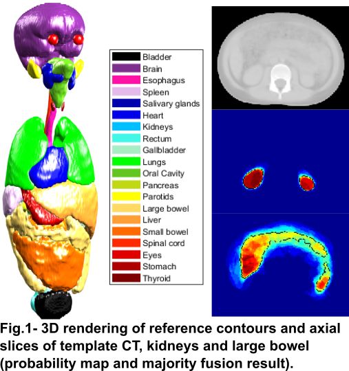
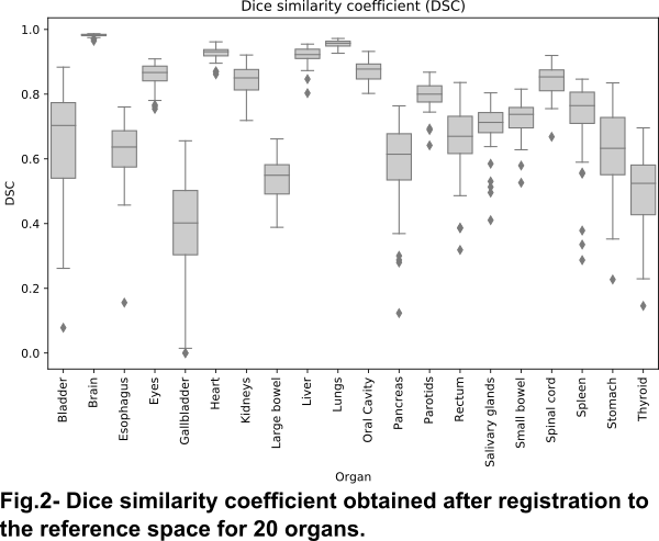
A reference space representative of the paediatric radiotherapy population, generated in a recent study, was used for spatial normalisation. The reference CT was constructed using groupwise image registration of twenty craniospinal irradiation paediatric CT images (mean age: 8, range: 3–15y). Reference contours were generated by majority fusion after co-registration, followed by simple post-processing operations. To comprehensively evaluate the constructed paediatric atlas for spatial normalisation at the whole-body level, an independent dataset of 54 craniospinal CTs (mean age: 8, range: 1–17y) with matching contours were deformably registered to the reference CT using NiftyReg. The segmentations consisted of 20 OAR associated with acute and late side-effects of treatment and were manually segmented by a single clinical expert on all datasets. The accuracy achieved for the inter-subject registration at the whole-body level was assessed by calculating the dice similarity coefficient (DSC) between deformed and reference segmentations.
Results
The reference CT and contours are shown in Fig.1. The anatomical shape and position was anatomically plausible, but probability maps were more diffuse for organs in the gastrointestinal tract, particularly smaller organs (e.g.: gallbladder), indicating larger interpatient anatomical variability for these organs. Fig. 2 shows the distribution of DSC values achieved for each individual contour on the independent dataset. The average DSC ranged from 0.384±0.164 (gallbladder) to 0.982±0.004 (brain), with average DSC larger than 0.7 for 12 of the 20 organs evaluated. The lower accuracy obtained in the remaining organs, mostly located in the abdomen, were likely due to larger inter-patient variability and poor contrast at soft tissue boundaries to guide the registrations, which hinders both the generation of the template contours and propagation of new subjects.
Conclusion
A previously developed atlas was shown to be an adequate common reference space for spatial normalisation for many organs of interest simultaneously, but performance must be improved for abdominal organs. Other label fusion approaches and more refined registration workflows should be investigated to increase mapping accuracy in small and/or highly deformable organs.