Copenhagen, Denmark
Onsite/Online

ESTRO 2022

Session Item

Radiomics, modelling and statistical methods
7011
Poster (digital)
Physics
Radiomic features are minimally repeatable in test-retest MR images of cervical cancer
Chelmis Muthoni Thiong'o, United Kingdom
PO-1781

Abstract

Radiomic features are minimally repeatable in test-retest MR images of cervical cancer
Authors:

Chelmis Thiong'o1, Alan McWilliam1, Gareth Price1, Angela Davey1, Andrew Green2

1University of Manchester, Division of Cancer Sciences, Manchester, United Kingdom; 2University of Manchester, Division of Cancer Sciences , Manchester, United Kingdom

Show Affiliations
Purpose or Objective

Radiomics promises to identify image characteristics that may predict treatment outcomes. However, identification of robust radiomic features is first required before building prognostic models. In this work, we performed a test-retest analysis to investigate repeatability of radiomic features in cervical cancer from T2-weighted (T2W) and Dixon MR images for the GTV and the peritumoral region.

Material and Methods

6 patients with FIGO stage IB2 and IIB cervical cancer had 3 MR scans each, with at least a week between scans. The scans were acquired on a single MR machine. At each scan, a T2-weighted image was taken at the start and end of the exam and Dixon sequences were sequentially acquired over 10 minutes. For this study, T2W images at the start and end of the scan and the first and last Dixon sequences in the scan were included, resulting in 4 images per scan. This selection results in a total of 36 test-retest pairs including both T2W and Dixon sequences.

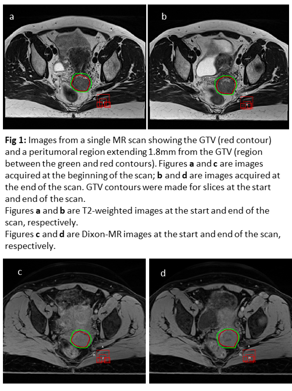
The GTV was contoured by a single observer on the start and end T2W images, and transferred through rigid registration to the start and end Dixon sequence. For each image, radiomic features were extracted from 2 regions: the GTV and a peritumoral region which extends from the GTV surface to 1.8mm (using the start GTV as reference). The regions are shown in Figure 1.

PyRadiomics was used to extract first-order, shape, and texture radiomic features from the original T2W and Dixon images, and their Laplacian of Gaussian-filtered (with σ = 3, 4, and 5) and Wavelet-filtered counterparts. This resulted in 1127 features per image.

Spearman’s ⍴ was used to exclude features highly correlated with volume (|⍴| ≥ 0.9). Of the remaining features, those with minimal variation between the start and end of a single scan were selected as being stable using Intraclass Correlation Coefficient, ICC (1,1) ≥ 0.9. Spearman’s ⍴ was then used to identify features that were not correlated with each other, with features having |⍴| < 0.65 being considered acceptable.


Results

In the GTV region, only original shape SurfaceVolumeRatio and MeshVolume features were repeatable in T2W and Dixon images. Original first-order InterquartileRange feature was also repeatable in T2W images.

In the peritumoral region, only original shape Elongation was repeatable in both T2W and Dixon images.

Figure 2a shows the number of radiomic features remaining at each step of the feature selection process. Less than 1% of radiomic features were stable and contained non-redundant information for potential prognostic modelling. Figure 2b shows Venn diagrams of identified stable features in the GTV and peritumoral regions for both T2W and Dixon sequences.


Conclusion

We show that features stable for one MR sequence are not necessarily stable in other sequences, and that the image region from which they are extracted can impact feature repeatability. We therefore advise region and modality specific test-retest analyses for selection of radiomic features for prognostic models.