MRI Radiomics in prostate cancer: a reliability study
PO-1778
Abstract
MRI Radiomics in prostate cancer: a reliability study
Authors: Antonio Angrisani1, Luca Mastrandrea1, Angelo Sangiovanni1, Valerio Nardone1, Roberta Grassi1, Alfonso Reginelli2, Cesare Guida3, Salvatore Cappabianca4
1“L. Vanvitelli” University of Campania, Precision Medicine - Radiotherapy Unit, Naples, Italy; 2"L. Vanvitelli" University of Campania, Precision Medicine - Radiology Unit, Naples, Italy; 3Ospedale del Mare, Radiotherapy, Naples, Italy; 4"L. Vanvitelli" University of Campania, Precision Medicine - Radiotherapy Unit, Naples, Italy
Show Affiliations
Hide Affiliations
Purpose or Objective
Radiomics
can provide quantitative features from medical imaging that can be correlated
to clinical endpoints. The challenges relevant to the robustness of radiomics
features have been analyzed by many researchers, as it seems to be influenced by
acquisition and reconstruction protocols, as well as by the segmentation of the
region of interest (ROI). Prostate cancer (PCa) represents a difficult playground for
this technique, due to the discrepancies in the identification of the cancer lesion
and the various acquisition protocols. The aim of this study is to investigate the reliability
of radiomics in prostate cancer detection.
Material and Methods
A homogeneous
cohort of patients, with a prostate-specific antigen (PSA) rise that underwent multiparametric MRI imaging
of the prostate before prostate biopsy, was tested in this study. All the patients were
acquired with the same MRI scanner, with a standardized protocol. The
identification of an MRI suspicious cancer lesion was done by two Radiologists
with great experience in prostate cancer (>10 years). The segmentation of
the lesion was done by two Residents (in Radiation Oncology and Radiologist). After the
segmentation, texture features were extracted with LIFEx software. All the patients
underwent random prostate biopsy procedures and the presence of prostate
cancer, as well as the Gleason score, was retrospectively collected. Texture features were then tested with intraclass coefficient
correlation (ICC) analysis to analyze the reliability of the segmentation.
Results
Forty-four
consecutive patients with suspect PCa were included in the present analysis. In 26 patients
(59,1%) the prostate biopsy confirmed the presence of PCa, which was
scored as Gleason 6 in 6 patients (13,6%), Gleason 3+4 in 8 patients (18,2), and
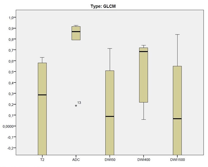
Gleason 4+3 in 12 patients (27.3%). The blind analysis conducted by two physicians as well as the ICC distribution led us to consider the ADC and the DWI400 sequences as the most reliable sequences. There were
significant differences in the distribution of ICC in GLCM features (p:0,018),
with no differences in the other subsections of Shape features (p:0,893), GLRLM
(p:0,594), NGLDM (p:0,109), GLZLM (p:0,594). The
reliability analysis, conversely, showed poor results in the majority of
the other MRI acquisitions (61% in T2, 89% in DWI50, 44% in DWI400, and 83% in
DWI1500), with ADC acquisition only showing better reliability. The ICC distribution of the GLCM features in five different acquisitions is shown in figure 1.
 .
.
Conclusion
The
low ratio of reliability in a monoinstitutional homogenous cohort represents a
significant alarm bell for the application of MRI Radiomics in the field of
prostate cancer. Supplementary work is needed in a clinical setting to further study the
potential of MRI radiomics in PCa.