A dosimetric evaluation of 4D-CT vs 3D-CT in mid and lower third upper GI Patients
Bethan Stewart-Thomson,
United Kingdom
PO-1293
Abstract
A dosimetric evaluation of 4D-CT vs 3D-CT in mid and lower third upper GI Patients
Authors: Bethan Stewart-Thomson1
1The Clatterbridge Cancer Centre, Physics, Liverpool, United Kingdom
Show Affiliations
Hide Affiliations
Purpose or Objective
This study will investigate whether patients
with a middle or lower-third oesophageal tumour will benefit from lower organ
at risk (OAR) doses with a 4D-CT planning scan compared with a 3D-CT planning
scan. The SCOPE studies have previously highlighted the benefits of a 4D-CT for
patients with conformal treatment plans. This study aims to determine if this
benefit extends to middle-third oesophageal patients and, whether there is an
additional benefit with using a more precise planning method of intensity-modulated radiotherapy (IMRT) such as volumetric modulated arc therapy (VMAT).
Material and Methods
At our centre, 3D scans are acquired
during free-breathing, without any monitoring of a patients breathing cycle. A
4D scan requires a regular breathing cycle, which is monitored prior to scan
acquisition to determine the patient’s suitability; this ensures the patient is
able to take regular, consistent breaths for the duration of treatment delivery.
Patients included in this study had previously been dual-scanned in 3D and 4D
and clinical plans were created using the 4D-CT scan, taking breathing motion
into account. At our centre, 4D-CT patients are treated with a reduced PTV
margin (0.5cm vs 1.0cm for 3D-CT). For this study, the 3D and 4D datasets were
anonymised, and the 3D scan was contoured by one of three clinicians, the 4D
scan having been contoured previously. New treatment plans were created
retrospectively for the 3D and 4D scans separately which allowed dosimetric
comparison of OAR doses and target volumes.
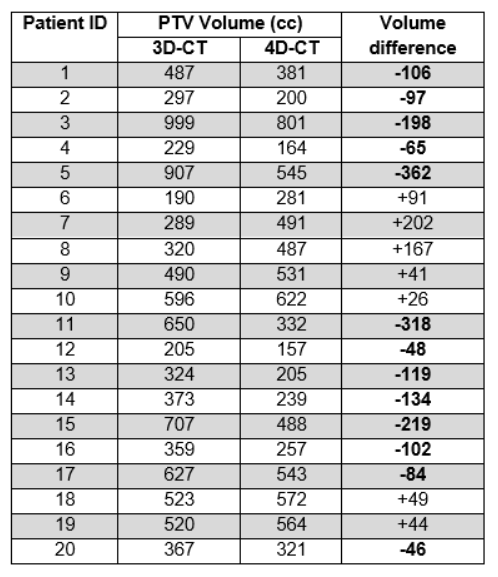
Results
Analysis of the results shows there
is a dosimetric benefit to planning with 4D-CT scans for both middle and
lower-third oesophagus patients with significant reductions in heart, lung and
spinal canal doses – as shown in table 1. In addition to this, there is a
reduction in overall target volume size using a 4D-CT – as shown in table 2.
Further research is required with a larger sample size to determine if these
results can be replicated.
table 1
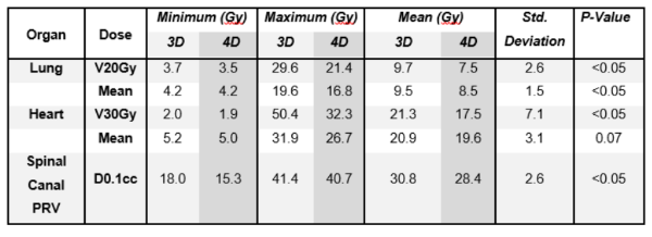
table 2
Conclusion
There is a statistically significant
benefit for patients with a middle or lower third oesophageal tumour to have a
4D-CT acquired for radiotherapy planning to reduce PTV volume and OAR doses. This
study has shown the dosimetric benefit of reducing OAR doses using VMAT with
the acquisition of a 4D-CT compared with a 3D-CT. This research has been
expanded by including patients with a middle third oesophageal tumour. The data
from this study can be used to justify the use of 4D-CTs for middle-third
oesophageal patients.