Copenhagen, Denmark
Onsite/Online

ESTRO 2022

Session Item

Monday
May 09
14:15 - 15:15
Poster Station 2
22: Gynaecological
Gabriella Macchia, Italy
3420
Poster Discussion
Clinical
Coverage probability planning is safe for simultaneously integrated boost of lymph nodes in c.vulva
Sandy Mohamed, Egypt
PD-0903

Abstract

Coverage probability planning is safe for simultaneously integrated boost of lymph nodes in c.vulva
Authors:

Sandy Mohamed1, Marianne S Assenholt2, Lars Fokdal3, Jesper Kallehauge4, Jacob C. Lindegaard3, Kari Tanderup5

1Cairo University, NCI, Department of Radiation Oncology, Cairo, Egypt; 2Aarhus University Hospital, Department of Medical Physics, Aarhus, Denmark; 3Aarhus University Hospital, Department of Oncology, Aarhus, Denmark; 4Danish Centre of particle therapy, Medical physics, Aarhus, Denmark; 5Aarhus University, Institute of Clinical Medicine, Aarhus, Denmark

Show Affiliations
Purpose or Objective

Pathological inguinal lymph nodes (PILN) are found in >50% of patients with vulvar cancer referred for radiotherapy (RT). Strategies for minimizing irradiation of organs at risk (OAR) from PILN boosting are needed to minimize the risk of morbidity arising in the groin such as lymphedema. Coverage probability (CovP) is a new planning strategy for simultaneous integrated boost (SIB). CovP mirrors the probabilities of nodal movement and regression during irradiation with a slight overdose in the nodal center and cooling of the PTV edge, allowing for a steep dose gradient towards OAR (Fig 1). Our aim was to investigate if SIB of PILN using CovP can be delivered safely in vulvar cancer

Material and Methods

Ten consecutive patients treated with definitive RT including SIB of PILN and with daily cone beam CT (CBCT) for all fractions (fx) were included. RT was delivered by VMAT. Dose to the elective target was 51.2/32 fx with a SIB of 64 Gy/32 fx to the primary tumor and PILN.

PILN were contoured on both planning CT and MRI (GTV-N). The images were rigidly registered. For each PILN, the GTV-N contoured on MRI and CT were combined to form ITV-N. Each PILN was also contoured on every second or third CBCT for a total of 11 CBCTs evaluated in all patients.

For OAR the following contours were considered: subcutaneous tissue (SC), inguinal vessels, skin rim, bowel and body contour.

Three plans were created for every patient:

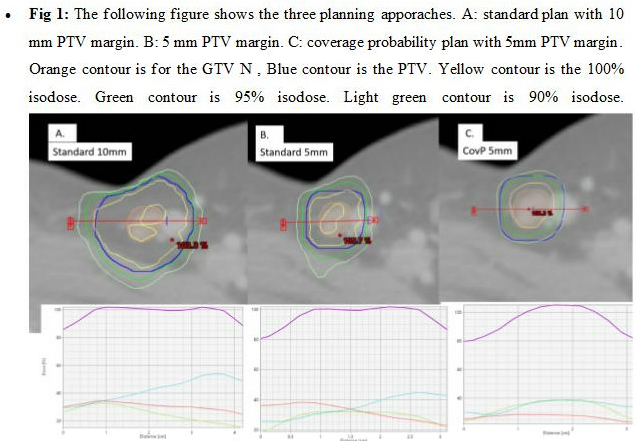
A– Standard CT based planning; PTV-N using GTV-NCT with a 10mm isotropic margin. GTV-NCT was delineated on the planning CT. The coverage criteria were PTV-N D98% ≥ 95% of prescribed dose (PD).

B– CT and MRI based planning with smaller margins: PTV-N using a 5mm isotropic margin added to ITV-N. The coverage criteria was the same as in plan A.

C– CovP: PTV-N was the same as in plan B. Coverage criteria were: ITV-N D98% ≥ 100% PD and PTV-N D98% ≥ 90% of PD.(Fig1).

Rigid registration was performed between CBCTs and the planning CT, and all the CBCTs delineations were propagated to the planning CT. For every PILN (contoured on the planning CT and each contoured CBCT: D98%, D50%, D2% were extracted for the GTV N on the CBCT and the PTV for the three plans. The total delivered dose was estimated by accumulating dose across all CBCT. Doses to OAR were evaluated on the planning CT for the three different planning approaches.


Results

Thirty-five PILNs were boosted in the included patients. There was no significant difference in accumulated D98% delivered to the GTV-N between the three plans. CovP based SIB delivered a higher mean dose to the GTV-N D50% (by about 2.5Gy) and D2% (by about 3Gy) compared to both plan A and B (p<0.01) (Table 1)

The planned mean doses to the OAR were reduced when applying CovP (Table 1). There was a significant reduction of the SC V55 when comparing CovP plan B strategy (p<0.001).

Conclusion

SIB of PILN in vulvar cancer based CovP and a 5mm PTV margin does not impede target coverage during long course RT and reduce the boost dose to normal tissues in the groin.