Copenhagen, Denmark
Onsite/Online

ESTRO 2022

Session Item

Sunday
May 08
10:30 - 11:30
Room D1
Lung
Cécile Le Péchoux, France;
Peter van Rossum, The Netherlands
2230
Proffered Papers
Clinical
10:50 - 11:00
Geometric and dosimetric stability of high FDG-uptake volumes during dose escalated RT of NSCLC
Ditte Sloth Møller, Denmark
OC-0439

Abstract

Geometric and dosimetric stability of high FDG-uptake volumes during dose escalated RT of NSCLC
Authors:

Ditte Sloth Møller1, Lone Hoffmann1, Ate Haraldsen2, Marianne Marquard Knap3, Azza Ahmed Khalil3, Christina Maria Lutz1

1Aarhus University Hospital, Department of Medical Physics, Aarhus, Denmark; 2Aarhus University Hospital, Department of Nuclear Medicine & PET-Centre, Aarhus, Denmark; 3Aarhus University Hospital, Department of Oncology, Aarhus, Denmark

Show Affiliations
Purpose or Objective

In the multi-centre phase III trial NARLAL2 (NTC NCT02354274) locally advanced NSCLC patients are randomised between a homogenous dose to the entire target (66Gy/33fx) and a heterogeneous dose escalation driven by the high FDG-uptake volumes (mean doses up to 95Gy/33fx). In this study, we investigate the geometric and dosimetric stability of these volumes.

Material and Methods

In total, 68 patients were included. They all had three repeated PET/CT scans at the same scanner: PET/CT0 (2 weeks before RT start, used for planning), PET/CT1 (1 week after RT start) and PET/CT2 (2 weeks after RT start). The GTV of the primary tumour was delineated on each scan. Within each GTV, the sub-volume with FDG-signal above a threshold of 50% of SUVpeak was segmented (V0, V1 and V2). CT1 and CT2 were rigidly registered to CT0 prioritizing the GTV area. The overlap fraction (OF) was defined as: OF0X=(V0∩VX)/min(V0,VX), X=1,2. Regardless of randomisation result, the dosimetric impact of changes in FDG uptake was investigated by recalculation of the escalated dose plan on CT2 (with V2). The resulting change in mean dose to high uptake areas V0 and V2 (ΔDV0,V2) was evaluated with respect to the cause of change. A Chi-square test was used to test if a low overlap OF02 (<0.7) was correlated with a large decrease in dose ΔDV2-V0 (>3Gy).

Results

The geometric stability of the high uptake volumes was high, with median (min;max) values of OF01 : 0.83 (0.42;1.00)  and OF02 :  0.82 (0.38;1.00). OF01 and OF02 for all patients are shown in fig1. 


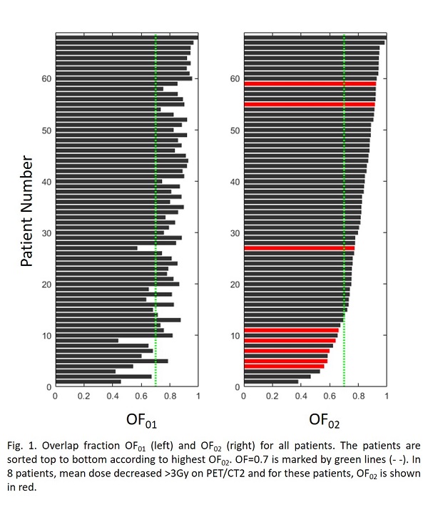
In 56 (82%) and 55 (80%) of the patients, the OF was above 0.7 at PET/CT1 and PET/CT2, respectively. Both increase (negative ΔDV0,V2) and decrease (positive ΔDV0,V2) in dose to V2 compared to V0 were 
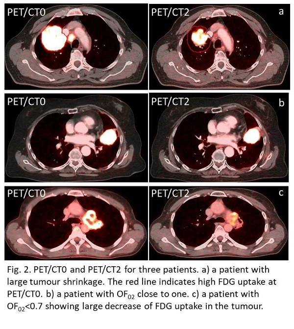
observed. Increase was in general due to tumour shrinkage as illustrated in fig2a. There was a significant correlation between patients with OF02<70% and patients with mean dose decrease >3Gy (p<0.001). Patients with >3Gy decrease are highlighted in fig1. For patients with OF02 above 0.7, the median change in dose, ΔDV0,V2, was 0.2Gy(-4.4;5.5), and in only 5% of these patients (3/55) the dose decrease >3Gy. For the 13 patients where the OF02 was below 0.7, the median change in dose, ΔDV0,V2, was 2.1Gy(-0.9;17.0), and in 38% of these patients (5/13) the dose decreased >3Gy.

The primary cause for OF02<0.7 was the decline of SUVpeak to ~2 times background activity, as this resulted in large changes of the region included in V2 for 54% of patients (7/13), see example in Fig 2c. Additionally, OF02<0.7 was observed for patients with larger anatomical changes, such as necrotic regions partly dissolved and changing atelectasis, where plan adaptation was warranted. 


Conclusion

For the majority of patients, the geometric and dosimetric stability of the high FDG-uptake volumes was high. For patients with OF02 below 0.7 a decrease of escalation dose of more than 3 Gy was more likely and mainly due to large decrease in SUVpeak or major anatomical changes.