Copenhagen, Denmark
Onsite/Online

ESTRO 2022

Session Item

Saturday
May 07
16:55 - 17:55
Auditorium 15
Haematology
Jessica Brady, United Kingdom;
Lena Specht, Denmark
1560
Proffered Papers
Clinical
16:55 - 17:05
Patterns of failure in primary CNS lymphoma: the MSKCC experience from 2004-2018
Kathryn Tringale, USA
OC-0291

Abstract

Patterns of failure in primary CNS lymphoma: the MSKCC experience from 2004-2018
Authors:

Kathryn Tringale1, Christian Grommes2, Behroze Vachha3, Harper Hubbeling1, Ari N. Wijetunga1, Carla Hajj1, Joachim Yahalom1, Brandon Imber1

1Memorial Sloan Kettering Cancer Center, Radiation Oncology, New York, USA; 2Memorial Sloan Kettering Cancer Center, Neuro-Oncology, New York, USA; 3Memorial Sloan Kettering Cancer Center, Radiology, New York, USA

Show Affiliations
Purpose or Objective

Treatment of primary CNS lymphoma (PCNSL) has rapidly evolved, emphasizing improved efficacy while reducing toxicity. Despite advances, many patients still relapse after first-line therapy. Clinical predictors and patterns of failure are critical to optimize and personalize induction and consolidation regimens and have been poorly studied in the modern era. Our objective was to evaluate PCNSL patterns of failure after contemporary treatment.

Material and Methods

PCNSL patients treated at Memorial Sloan Kettering Cancer Center between 2004-2018 were evaluated. Initial site(s) of T1 post-contrast-enhancing disease on baseline MRI was characterized by laterality, uni- vs multifocality, supra- vs infratentorial, and deep nuclei, CSF, and/or radiographic meningeal involvement. For patients who received consolidation, disease status (radiographic evidence of contrast-enhancing disease [EOD] vs no evidence of disease [NED]) was evaluated on pre-consolidation MRI. Site of post-induction relapse was characterized as local (involving or adjacent to initial site) vs distant intracranial. Radiographic predictors of relapse patterns were described with univariable logistic regression. Time-to-failure (TTF) and overall survival (OS) analyses were performed using Kaplan Meier with log-rank.

Results

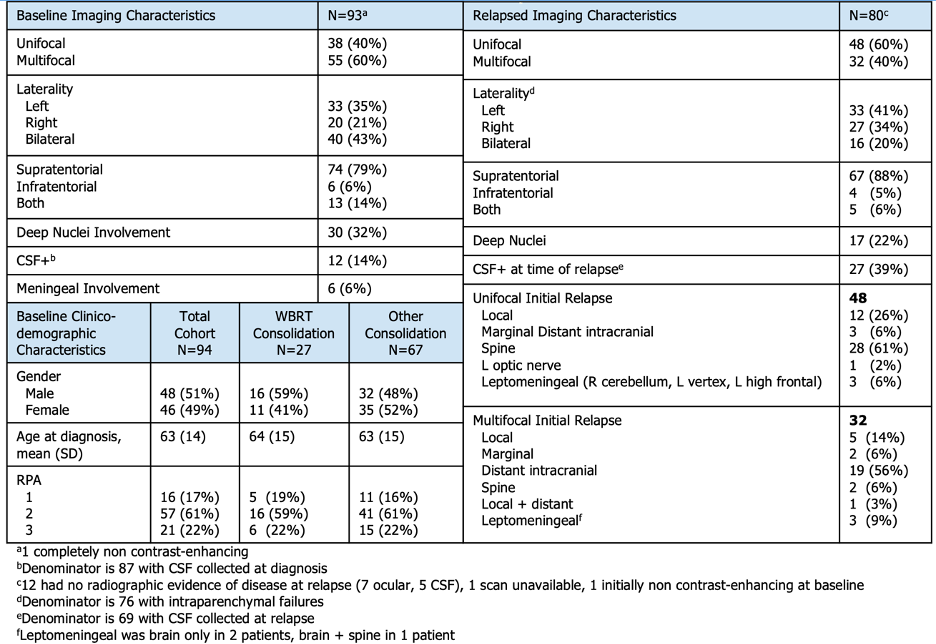
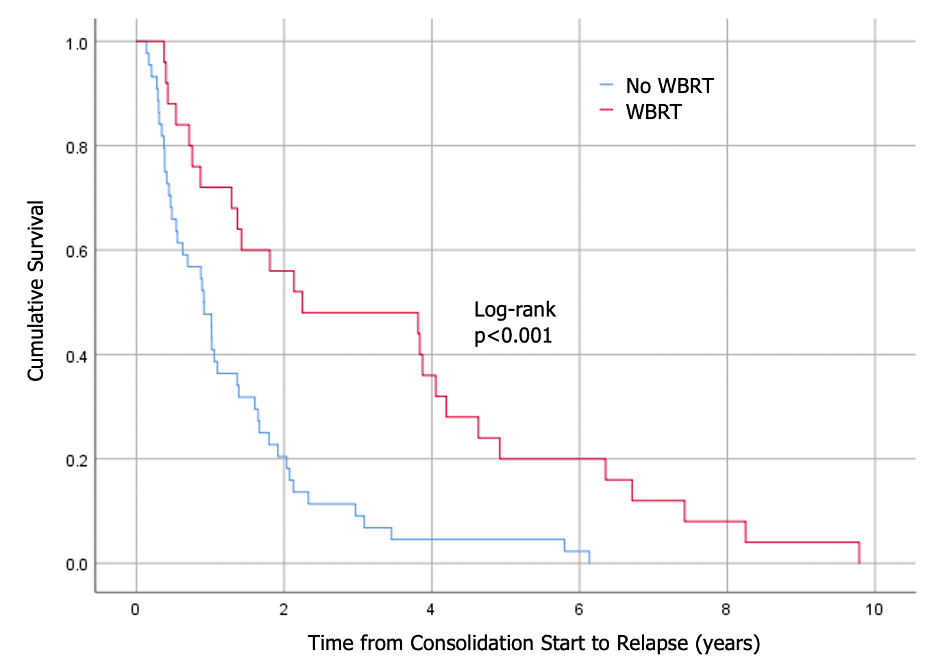
Of 259 patients treated with median follow-up of 3.7 years (range: 0.4-11.0), 94 (36%) relapsed at a median time of 1.3 years (range: 0.1-10.1) from induction initiation. Most received induction R-MVP (n=66, 70%) and most were consolidated (n=75, 80%), with cytarabine (n=42, 56%) or whole brain radiotherapy (WBRT, n=27, 29%). At baseline, most had multifocal (n=55, 60%) and supratentorial disease (n=74, 79%; Table). Fewer involved deep nuclei (n=30, 32%), meninges (n=6, 6%), or CSF (n=12 of 87 collected, 14%). At relapse, 80 (85%) had radiographic disease, 12 (13%) had cytologic evidence only (7 ocular on biopsy, 5 CSF involvement), and 2 (2%) scans were not evaluable. Of 80 radiographic relapses, most were supratentorial (61, 80%) and maintained laterality (17 of 33 [59%] ipsilateral left, 11 of 20 [55%] ipsilateral right). Intracranial failures (n=76) were mostly distant (n=54, 71%) with fewer relapsing both locally and unifocally (n=15, 20%). Patients with EOD pre-consolidation were more likely to have a local failure (OR 4.8 [95% CI 1.1-7.1], p=0.06). WBRT consolidation was associated with longer TTF (2.2 vs 0.9 years, p<0.001; Figure) and delivered mostly (n=23, 85%) with reduced dose (2340cGy/13fx). Median OS trended toward improved with WBRT consolidation (7.4 vs 4.2 years, p=0.056).


Conclusion

While intracranial PCNSL relapses were often distant from the site of initial disease, most maintained laterality and EOD pre-consolidation predicted local failure. Consolidation with WBRT (mostly with reduced dose regimen) was associated with prolonged TTF and trended toward improved OS, supporting results from NRG-RTOG 1114. Future analyses will evaluate predictors of relapse.