Enhanced RBE for late compared to early normal tissue damage in vivo
Cathrine Bang Overgaard,
Denmark
OC-0092
Abstract
Enhanced RBE for late compared to early normal tissue damage in vivo
Authors: Cathrine Bang Overgaard1, Brita Singers Sørensen2, Niels Bassler3, Per Poulsen4, Cai Grau3, Jens Overgaard1, Mateusz Krzysztof Sitarz3, Harald Spejlborg5
1Aarhus University Hospital, Experimental Clinical Oncology, Aarhus, Denmark; 2Aarhus University, Danish Centre for Particle Therapy, Aarhus, Denmark; 3Aarhus University Hospital, Danish Centre for Particle Therapy, Aarhus, Denmark; 4Aarhus University Hospital , Danish Centre for Particle Therapy, Aarhus, Denmark; 5Aarhus University Hospital, Oncology, Aarhus, Denmark
Show Affiliations
Hide Affiliations
Purpose or Objective
Protons deliver the highest physical dose in a specific-targeted tissue area. This feature makes protons compared to x-rays, in some cases, a more favorable radiation modality to use in treatment of different cancers. Proton radiation has an increased relative radiobiological effectiveness (RBE) compared to X-rays. Despite that RBE is not constant and depends on factors such as tissue type, dose per fractions, position in the Spread-Out Bragg Peak (SOBP) and endpoint, a constant RBE value of 1.1 is used clinically. Several in vitro studies have been conducted within the area of cell response, but in vivo RBE data are still scarce. In the current study both acute and late normal tissue response are analyzed in the same animals to assess the RBE in vivo.
Material and Methods
Included in the study were 243 12–14-week-old C3H/HeNRj mice that were fixated in jigs and placed on top of a heated (25°C) water phantom. Their right hind leg was submerged into the water phantom and placed within a well-defined radiation field. Single doses of 22-46 Gy were delivered in the center of a SOBP by an 83-107MeV pencil beam scanning proton beam. As reference, a 6 MV photon beam from a clinical linear accelerator was used.
Early and late normal tissue damages were investigated by two endpoints: (1) acute moist desquamation of the skin within 30 days post irradiation, and (2) radiation induced fibrosis developed within 9 to 53 weeks post irradiation.
Results
The late effect ED50 (the dose producing radiation induced fibrosis in 50% of the mice) were 31.63 Gy (29.72 Gy-33.34 Gy) for protons and 38.26 Gy (34.44 Gy-50.21 Gy) for photons indicating a significant higher ED50 for protons (p=0.035).
No significant difference was found between protons and photons for the acute skin damage. The corresponding RBE values were 1.05 (1.00-1.10; moist desquamation) and 1.21 (1.05-1.36; fibrosis) (see figure 1).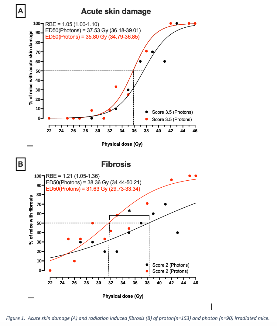
Conclusion
The data indicate that proton radiation leads to a higher RBE for late reacting endpoints compared to early reacting endpoints analyzed in the same tissue. This is in agreement with data from in vitro cell survival curves, suggesting late reacting endpoints to display higher RBE values than early reacting endpoints. There is a need for more research into the late effects of proton radiation.