Investigation of obstructions in ring applicators during pulsed dose rate cervix brachytherapy
PO-0214
Abstract
Investigation of obstructions in ring applicators during pulsed dose rate cervix brachytherapy
Authors: Geetha Menon1, Brent Long2, Rebecca Petit3, Janet Zimmer3, Kimberly Gadbois3, Yury Niatsetski4, Ericka Wiebe1, Julie Cuartero1, Fleur Huang1, Eugene Yip1
1Cross Cancer Institute & University of Alberta, Oncology, Edmonton, Canada; 2Cross Cancer Institute, Medical Physics, Edmonton, Canada; 3Cross Cancer Institute, Radiation Therapy, Edmonton, Canada; 4Elekta, Physics and Advanced Development , Veenendaal, The Netherlands
Show Affiliations
Hide Affiliations
Purpose or Objective
During intracavitary pulsed dose rate (PDR) brachytherapy (BT) for
cervical cancer using ring applicators, intermittent friction or obstruction
errors are encountered with the check and source cables, requiring operator intervention.
This study ascertains patterns and plausible causes of these errors by
analyzing the actual source paths in the rings.
Material and Methods
Errors from 60 consecutive 58-hourly-pulse BT treatments using
Interstitial CT/MR ring applicators on 2 microSelectron PDR afterloaders
(Elekta, Sweden) were identified. We evaluated obstruction patterns caused by the
check and source cables during both out- and in-drives. To determine the Ir-192
source paths in the rings (diameters of 26 and 30 mm), a portable x-ray machine
(GE AMX-4, GE Healthcare, USA) was used to image the source capsule (Fig. 1(a))
at pre-programmed dwell positions: (i) 6 dwells at 15 mm separation (Fig 1(b); illustrates
complete path in ring) and (ii) 5-6 dwells at 5 mm separation (Fig 1(c); illustrates
capsule position at adjacent dwells). The check cable path was fluoro-imaged
when the capsule reached the distal ring end; however, the cable’s high travel
speed precluded images suitable for drawing conclusions. Repeated runs were
performed with the transfer tube in different arrangements (alignment,
curvature) relative to the ring to reproduce the obstructions.

Results
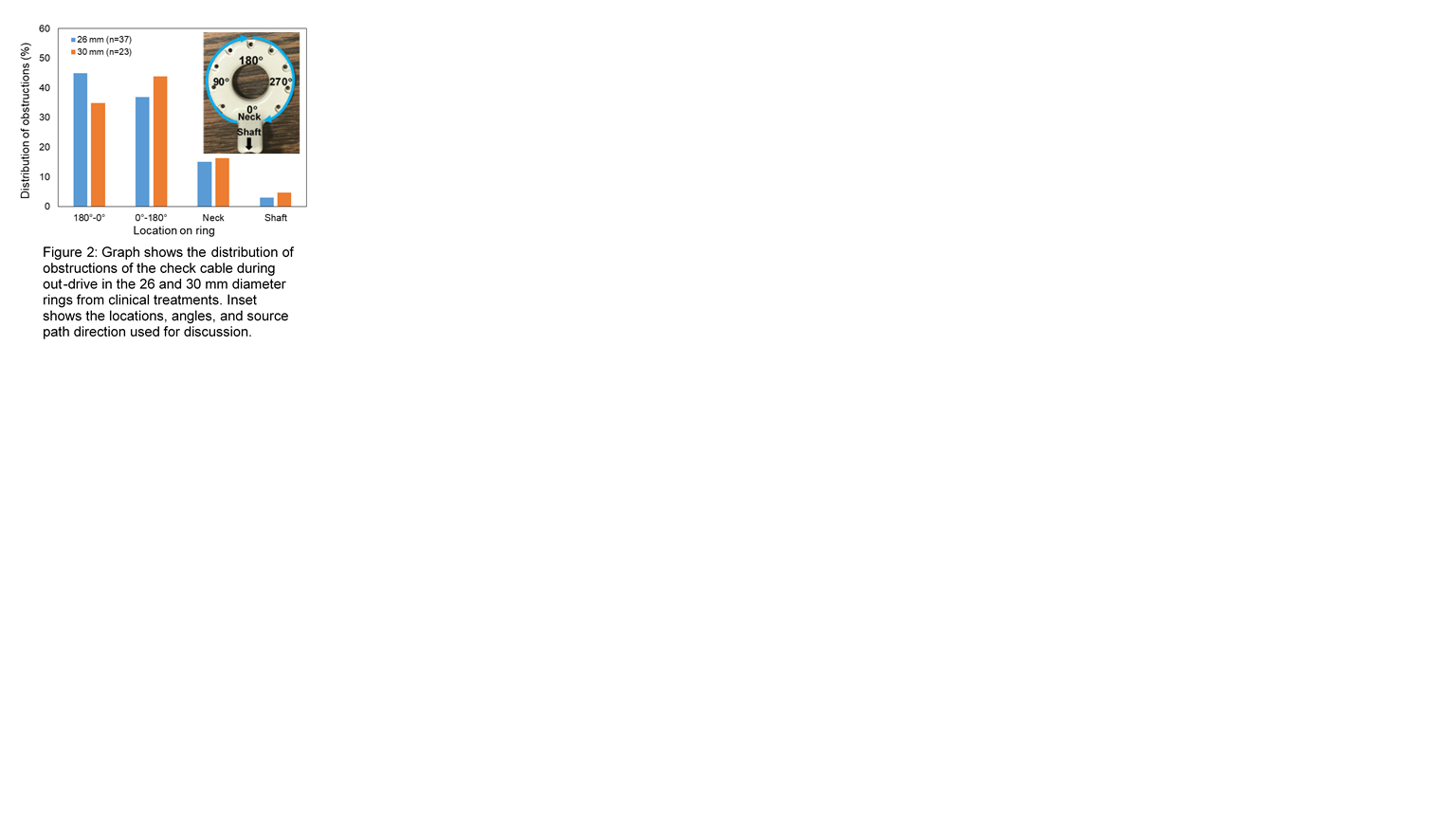
In 45 of 60 ring treatments (26 mm (n=37)
and 30mm (n=23), the check cable produced in total 625 obstructions, mostly in the
curved portion of the ring (81.9% for 26mm (54.9% from 180°-0°) and 78.8% for 30mm (55.7% from 0°-180°)) during out-drive. Only 2 obstructions
arose during in-drive. Image analysis showed that the source capsule tip comes
quite close to the outer wall of the lumen (width ~ 3.0mm for all rings) near 90° and 270° (Fig 2). With greater drive power for
the source cable (~ 10% more), fewer obstructions were encountered: 24 during out-drive
and none during in-drive. 15.1% (26mm) and 16.4% (30mm) of obstructions during
check cable out-drive occurred at the neck where the source enters the curved ring
from a straight path in the shaft. None of the obstructions were reproducible during
repeated runs with the transfer tube aligned with the ring. However, with curving
of the transfer tube (as can be seen in clinical PDR setups), occasional obstructions
could be generated, but only for the 26mm ring, demonstrating the importance of
transfer tube alignment with the rings during treatment.

Conclusion
Obstruction errors in cervix BT may be mitigated
by delivery in lithotomy setup and ideal transfer tube alignment with the ring,
more easily achievable in high-dose-rate BT settings. For PDR treatments with
multiple hourly in- and out-drives in the ring, and patient movement
during/in-between treatment, frequent transfer tube inspections to maintain path
of least curvature between the ring and afterloader indexer head should be
performed, particularly for smaller ring sizes.