Practical considerations on the use of EM tracking technology for clinical HDR brachytherapy
PO-0209
Abstract
Practical considerations on the use of EM tracking technology for clinical HDR brachytherapy
Authors: Marie Claude Lavallee1, Audrey Cantin2, Eric Vigneault2, William Foster2, Sylviane Aubin2, André-Guy Martin3, Martine Lefebvre2, Luc Beaulieu2
1CHU de quebec, radiation oncology, Quebec, Canada; 2CHU de Quebec, radiation oncology, Quebec, Canada; 3CHU de Quebec, Radiation oncology, Quebec, Canada
Show Affiliations
Hide Affiliations
Purpose or Objective
The electromagnetic tracking (EMT) system for intra-op US based HDR
brachytherapy is a new technology for tracking and automatic reconstruction of
catheters. The aim of this work is to
summarise the challenges pose by the technology and how to tackle them
Material and Methods
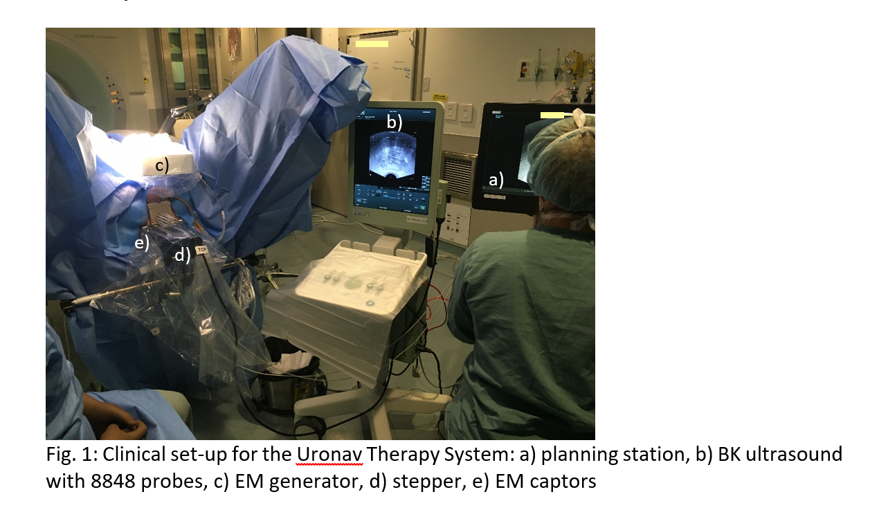
The Uronav Therapy system from Philips Disease Management Solutions was commissioned and integrated in our clinical practice (Fig.1). The calibration of the EMT to the US
image is a crucial step, and to obtain good results, a list of conditions
should be respected. The calibration validation must be done at the OR with the
same clinical set-up and in a US compatible prostate phantom. The clinical use
of that system also comes with special requirements starting with the patient
set-up, equipment used around the system, etc. Finally, organ delineation and
catheter’s tracking also come with challenges. Various solutions were explored
and are presented
Results
A critical step to ensure
accurate results is the registration of the EMT reference frame to the US. This
needs to be done in accordance to TG-128 (salty water - 43g/L). No metal part should be within
the EM field. A small metal rod hidden in our water container hinge introduced
an error up to 1.8mm. This further includes any support with metallic parts
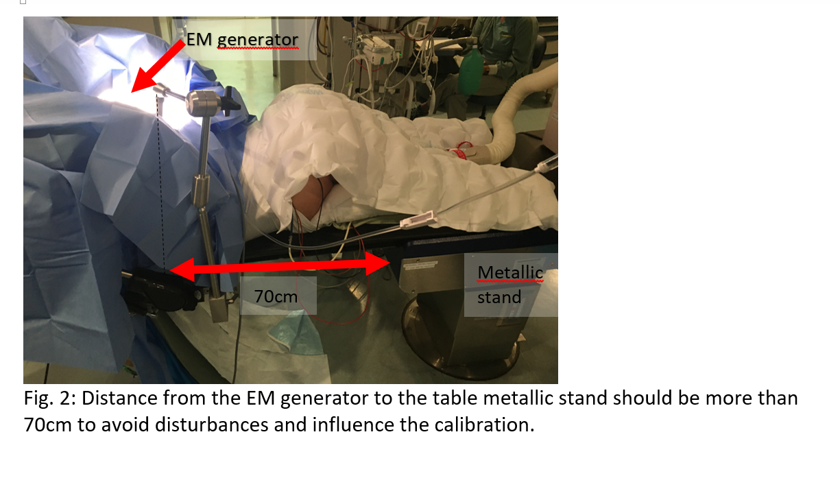
used to hold the calibration phantom. Another aspect is the clinical
environment. Metallic parts on the OR table can create EM disturbances and
introduce errors (Fig.2). In our set-up, the distance between the EM generator
and the table stand needs to be more than 70cm to avoid disturbance, which
translate in error of 2mm on tip positions and height of reconstructed
catheters. Cell phones, metal clamps and so on shouldn’t be placed close to the
EM field, as they introduce error relative to the calibration up to 2mm.
However, stirrups as well as surgical or vasectomy clips did not show any
effect. In this trial, no patient with pacemaker, neurostimulator, implanted
insulin pump, hip or knee prosthesis was allowed in order to avoid disturbance. Finally, the stylet used for EM tracking and automatic
reconstruction is not rigid enough to allow for an easy tissue perforation, but
stiffer than the source cable, sometimes introducing catheter motion during
retraction with an AP shift of the tip up to 3mm. Thus, it remains important to
visualize the reconstruction with sagittal live US imaging.
Conclusion
EM tracking offers the possibility of fast and
accurate solution for catheter guidance and reconstruction in US-guided
prostate HDR. Pointers were given beyond the vendor provided guidance to avoid
potential pitfalls and ensure that the stated accuracy is indeed reached